1NHK

CRYSTAL STRUCTURE OF MYXOCOCCUS XANTHUS NUCLEOSIDE DIPHOSPHATE KINASE AND ITS INTERACTION WITH A NUCLEOTIDE SUBSTRATE AT 2.0 ANGSTROMS RESOLUTION


Experimental Data Snapshot

  • Method: X-RAY DIFFRACTION
  • Resolution: 1.90 Å
  • R-Value Free: 0.206 
  • R-Value Work: 0.174 
  • R-Value Observed: 0.174 

wwPDB Validation   3D Report Full Report


Ligand Structure Quality Assessment 


This is version 1.3 of the entry. See complete history


Literature

The 1.9 A crystal structure of a nucleoside diphosphate kinase complex with adenosine 3',5'-cyclic monophosphate: evidence for competitive inhibition.

Strelkov, S.V.Perisic, O.Webb, P.A.Williams, R.L.

(1995) J Mol Biol 249: 665-674

  • DOI: https://doi.org/10.1006/jmbi.1995.0327
  • Primary Citation of Related Structures:  
    1NHK

  • PubMed Abstract: 

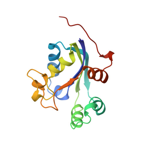
    The X-ray structure of Myxococcus xanthus nucleoside diphosphate (NDP) kinase complexed with adenosine 3',5'-cyclic monophosphate (cAMP) has been determined. The structure was solved by difference Fourier analysis. The refined structure has a crystallographic R-factor of 0.17 at 1.9 A resolution. The phosphoryl group and ribose moiety make extensive polar interactions with the protein, whereas the base interacts only with two hydrophobic residues. The comparison with the structure of the enzyme complex with the substrate adenosine diphosphate (ADP) reported earlier shows that cAMP and ADP interact similarly with the enzyme. The base of the cAMP is present in two conformations, syn and anti, with respect to the sugar. The syn conformer is dominant. Based on the effect of cAMP on phosphorylation of the human NDP kinase NM23, it had been proposed that cAMP might interact with NDP kinase in a manner distinct from other nucleotides. However, the structure of the M. xanthus NDP kinase/cAMP complex indicates that the nucleotide is a competitive inhibitor of the enzyme and occupies the usual nucleotide site. Kinetic assays of the NDP kinase activity in the presence of cAMP were done. Their results are consistent with a competitive character of the cAMP inhibition.


  • Organizational Affiliation

    Centre for Protein Engineering, Medical Research Council Centre, Cambridge, England.


Macromolecules
Find similar proteins by:  (by identity cutoff)  |  3D Structure
Entity ID: 1
MoleculeChains Sequence LengthOrganismDetailsImage
NUCLEOSIDE DIPHOSPHATE KINASEA [auth R],
B [auth L]
144Myxococcus xanthusMutation(s): 0 
Gene Names: CMP
EC: 2.7.4.6
UniProt
Find proteins for P15266 (Myxococcus xanthus)
Explore P15266 
Go to UniProtKB:  P15266
Entity Groups  
Sequence Clusters30% Identity50% Identity70% Identity90% Identity95% Identity100% Identity
UniProt GroupP15266
Sequence Annotations
Expand
  • Reference Sequence
Small Molecules
Ligands 1 Unique
IDChains Name / Formula / InChI Key2D Diagram3D Interactions
CMP
Query on CMP

Download Ideal Coordinates CCD File 
C [auth R],
D [auth L]
ADENOSINE-3',5'-CYCLIC-MONOPHOSPHATE
C10 H12 N5 O6 P
IVOMOUWHDPKRLL-KQYNXXCUSA-N
Experimental Data & Validation

Experimental Data

  • Method: X-RAY DIFFRACTION
  • Resolution: 1.90 Å
  • R-Value Free: 0.206 
  • R-Value Work: 0.174 
  • R-Value Observed: 0.174 
  • Space Group: P 43 21 2
Unit Cell:
Length ( Å )Angle ( ˚ )
a = 63.6α = 90
b = 63.6β = 90
c = 158.3γ = 90
Software Package:
Software NamePurpose
X-PLORmodel building
X-PLORrefinement
X-PLORphasing

Structure Validation

View Full Validation Report


Ligand Structure Quality Assessment 


Entry History 

Deposition Data

Revision History  (Full details and data files)

  • Version 1.0: 1995-03-31
    Type: Initial release
  • Version 1.1: 2008-03-10
    Changes: Version format compliance
  • Version 1.2: 2011-07-13
    Changes: Version format compliance
  • Version 1.3: 2024-02-14
    Changes: Data collection, Database references, Derived calculations, Other