1NHC | pdb_00001nhc

Structural insights into the processivity of endopolygalacturonase I from Aspergillus niger


Experimental Data Snapshot

  • Method: X-RAY DIFFRACTION
  • Resolution: 1.70 Å
  • R-Value Free: 
    0.209 (Depositor), 0.217 (DCC) 
  • R-Value Work: 
    0.175 (Depositor), 0.185 (DCC) 
  • R-Value Observed: 
    0.176 (Depositor) 

Starting Model: experimental
View more details

wwPDB Validation 3D Report Full Report

Validation slider image for 1NHC

Ligand Structure Quality Assessment 


This is version 3.0 of the entry. See complete history

Literature

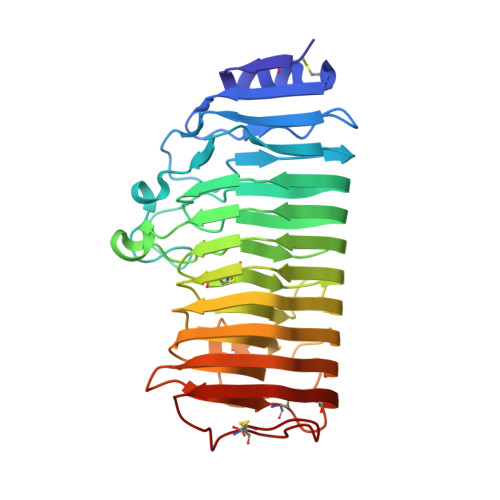
Structural insights into the processivity of endopolygalacturonase I from Aspergillus niger.

van Pouderoyen, G.Snijder, H.J.Benen, J.A.Dijkstra, B.W.

(2003) FEBS Lett 554: 462-466

  • DOI: https://doi.org/10.1016/s0014-5793(03)01221-3
  • Primary Citation Related Structures: 
    1NHC

  • PubMed Abstract: 

    Endopolygalacturonase I is a processive enzyme, while the 60% sequence identical endopolygalacturonase II is not. The 1.70 A resolution crystal structure of endopolygalacturonase I reveals a narrowed substrate binding cleft. In addition, Arg96, a residue in this cleft previously shown to be critical for processivity, interacts with the substrate mimics glycerol and sulfate in several well-defined conformations in the six molecules in the asymmetric unit. From this we conclude that both Arg96 and the narrowed substrate binding cleft contribute to retaining the substrate while it moves through the active site after a cleavage event has occurred.


  • Organizational Affiliation
    • Laboratory of Biophysical Chemistry, University of Groningen, Nijenborgh 4, 9747 AG Groningen, The Netherlands.

Macromolecule Content 

  • Total Structure Weight: 216.08 kDa 
  • Atom Count: 16,429 
  • Modeled Residue Count: 2,016 
  • Deposited Residue Count: 2,016 
  • Unique protein chains: 1

Macromolecules

Find similar proteins by:|  3D Structure
Entity ID: 1
MoleculeChains  Sequence LengthOrganismDetailsImage
Polygalacturonase I
A, B, C, D, E
A, B, C, D, E, F
336Aspergillus nigerMutation(s): 0 
EC: 3.2.1.15
UniProt
Find proteins for P26213 (Aspergillus niger)
Explore P26213 
Go to UniProtKB:  P26213
Entity Groups
Sequence Clusters30% Identity50% Identity70% Identity90% Identity95% Identity100% Identity
UniProt GroupP26213
Glycosylation
Glycosylation Sites: 3
Sequence Annotations
Expand
Reference Sequence

Oligosaccharides

Help  
Entity ID: 2
MoleculeChains Length2D Diagram GlycosylationD Interactions
2-acetamido-2-deoxy-beta-D-glucopyranose-(1-4)-2-acetamido-2-deoxy-beta-D-glucopyranose
G
2N-Glycosylation
Glycosylation Resources
GlyTouCan: G42666HT
GlyCosmos: G42666HT
GlyGen: G42666HT
Entity ID: 3
MoleculeChains Length2D Diagram GlycosylationD Interactions
alpha-D-mannopyranose-(1-3)-[alpha-D-mannopyranose-(1-6)]alpha-D-mannopyranose-(1-6)-beta-D-mannopyranose-(1-4)-2-acetamido-2-deoxy-beta-D-glucopyranose-(1-4)-2-acetamido-2-deoxy-beta-D-glucopyranose
H
6N-Glycosylation
Glycosylation Resources
GlyTouCan: G94106MV
GlyCosmos: G94106MV
GlyGen: G94106MV
Entity ID: 4
MoleculeChains Length2D Diagram GlycosylationD Interactions
alpha-D-mannopyranose-(1-4)-2-acetamido-2-deoxy-beta-D-glucopyranose-(1-4)-2-acetamido-2-deoxy-beta-D-glucopyranose
I
3N-Glycosylation
Glycosylation Resources
GlyTouCan: G62182OO
GlyCosmos: G62182OO
GlyGen: G62182OO

Small Molecules

Ligands 4 Unique
IDChains Name / Formula / InChI Key2D Diagram3D Interactions
NAG

Query on NAG


Download:Ideal Coordinates CCD File
HA [auth E],
OA [auth F],
V [auth C]
2-acetamido-2-deoxy-beta-D-glucopyranose
C8 H15 N O6
OVRNDRQMDRJTHS-FMDGEEDCSA-N
MAN

Query on MAN


Download:Ideal Coordinates CCD File
AA [auth D]
BA [auth D]
FA [auth E]
GA [auth E]
J [auth A]
AA [auth D],
BA [auth D],
FA [auth E],
GA [auth E],
J [auth A],
K [auth A],
NA [auth F],
O [auth B],
P [auth B],
T [auth C],
U [auth C]
alpha-D-mannopyranose
C6 H12 O6
WQZGKKKJIJFFOK-PQMKYFCFSA-N
SO4

Query on SO4


Download:Ideal Coordinates CCD File
CA [auth D]
DA [auth D]
IA [auth E]
JA [auth E]
KA [auth E]
CA [auth D],
DA [auth D],
IA [auth E],
JA [auth E],
KA [auth E],
L [auth A],
LA [auth E],
M [auth A],
PA [auth F],
Q [auth B],
QA [auth F],
R [auth B],
W [auth C],
X [auth C],
Y [auth C],
Z [auth C]
SULFATE ION
O4 S
QAOWNCQODCNURD-UHFFFAOYSA-L
GOL

Query on GOL


Download:Ideal Coordinates CCD File
EA [auth D],
MA [auth E],
N [auth A],
RA [auth F],
S [auth B]
GLYCEROL
C3 H8 O3
PEDCQBHIVMGVHV-UHFFFAOYSA-N

Experimental Data & Validation

Experimental Data

  • Method: X-RAY DIFFRACTION
  • Resolution: 1.70 Å
  • R-Value Free:  0.209 (Depositor), 0.217 (DCC) 
  • R-Value Work:  0.175 (Depositor), 0.185 (DCC) 
  • R-Value Observed: 0.176 (Depositor) 
Space Group: P 1
Unit Cell:
Length ( Å )Angle ( ˚ )
a = 68.012α = 114.32
b = 84.13β = 98
c = 96.035γ = 89.75
Software Package:
Software NamePurpose
DENZOdata reduction
TRUNCATEdata reduction
EPMRphasing
REFMACrefinement
CCP4data scaling

Structure Validation

View Full Validation Report


Ligand Structure Quality Assessment 


Entry History 

Deposition Data

Revision History  (Full details and data files)

  • Version 1.0: 2003-11-25
    Type: Initial release
  • Version 1.1: 2008-04-29
    Changes: Version format compliance
  • Version 1.2: 2011-07-13
    Changes: Non-polymer description, Version format compliance
  • Version 2.0: 2020-07-29
    Type: Remediation
    Reason: Carbohydrate remediation
    Changes: Advisory, Atomic model, Data collection, Derived calculations, Structure summary
  • Version 2.1: 2023-08-16
    Changes: Data collection, Database references, Refinement description, Structure summary
  • Version 3.0: 2024-12-25
    Changes: Advisory, Atomic model, Data collection, Derived calculations, Structure summary