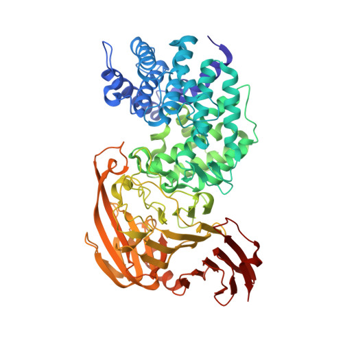
1N7R

Streptococcus pneumoniae Hyaluronate Lyase W291A/W292A/F343V Mutant complex with hexasaccharide hyaluronan


Experimental Data Snapshot

  • Method: X-RAY DIFFRACTION
  • Resolution: 2.20 Å
  • R-Value Free: 0.222 
  • R-Value Work: 0.177 
  • R-Value Observed: 0.177 

wwPDB Validation   3D Report Full Report


This is version 2.1 of the entry. See complete history


Literature

The function of hydrophobic residues in the catalytic cleft of Streptococcus pneumoniae hyaluronate lyase. Kinetic characterization of mutant enzyme forms

Nukui, M.Taylor, K.B.McPherson, D.T.Shigenaga, M.Jedrzejas, M.J.

(2003) J Biol Chem 278: 3079-3088

  • DOI: https://doi.org/10.1074/jbc.M204999200
  • Primary Citation of Related Structures:  
    1N7N, 1N7O, 1N7P, 1N7Q, 1N7R

  • PubMed Abstract: 

    Streptococcus pneumoniae hyaluronate lyase is a surface antigen of this Gram-positive human bacterial pathogen. The primary function of this enzyme is the degradation of hyaluronan, which is a major component of the extracellular matrix of the tissues of vertebrates and of some bacteria. The enzyme degrades its substrate through a beta-elimination process called proton acceptance and donation. The inherent part of this degradation is a processive mode of action of the enzyme degrading hyaluronan into unsaturated disaccharide hyaluronic acid blocks from the reducing to the nonreducing end of the polymer following the initial random endolytic binding to the substrate. The final degradation product is the unsaturated disaccharide hyaluronic acid. The residues of the enzyme that are involved in various aspects of such degradation were identified based on the three-dimensional structures of the native enzyme and its complexes with hyaluronan substrates of various lengths. The catalytic residues were identified to be Asn(349), His(399), and Tyr(408). The residues responsible for the release of the product of the reaction were identified as Glu(388), Asp(398), and Thr(400), and they were termed negative patch. The hydrophobic residues Trp(291), Trp(292), and Phe(343) were found to be responsible for the precise positioning of the substrate for enzyme catalysis and named hydrophobic patch. The comparison of the specific activities and kinetic properties of the wild type and the mutant enzymes involving the hydrophobic patch residues W292A, F343V, W291A/W292A, W292A/F343V, and W291A/W292A/F343V allowed for the characterization of every mutant and for the correlation of the activity and kinetic properties of the enzyme with its structure as well as the mechanism of catalysis.


  • Organizational Affiliation

    Children's Hospital Oakland Research Institute, Oakland, California 94609, USA.


Macromolecules
Find similar proteins by:  (by identity cutoff)  |  3D Structure
Entity ID: 1
MoleculeChains Sequence LengthOrganismDetailsImage
HYALURONIDASE721Streptococcus pneumoniaeMutation(s): 3 
EC: 4.2.2.1
UniProt
Find proteins for Q54873 (Streptococcus pneumoniae serotype 4 (strain ATCC BAA-334 / TIGR4))
Explore Q54873 
Go to UniProtKB:  Q54873
Entity Groups  
Sequence Clusters30% Identity50% Identity70% Identity90% Identity95% Identity100% Identity
UniProt GroupQ54873
Glycosylation
Glycosylation Sites: 1
Sequence Annotations
Expand
  • Reference Sequence
Oligosaccharides

Help

Entity ID: 2
MoleculeChains Length2D Diagram Glycosylation3D Interactions
beta-D-glucopyranuronic acid-(1-3)-2-acetamido-2-deoxy-beta-D-glucopyranose-(1-4)-beta-D-glucopyranuronic acid-(1-3)-2-acetamido-2-deoxy-beta-D-glucopyranose-(1-4)-beta-D-glucopyranuronic acid-(1-3)-2-acetamido-2-deoxy-beta-D-glucopyranose
B
6N/A
Glycosylation Resources
GlyTouCan:  G01866TT
GlyCosmos:  G01866TT
GlyGen:  G01866TT
Experimental Data & Validation

Experimental Data

  • Method: X-RAY DIFFRACTION
  • Resolution: 2.20 Å
  • R-Value Free: 0.222 
  • R-Value Work: 0.177 
  • R-Value Observed: 0.177 
  • Space Group: P 21 21 21
Unit Cell:
Length ( Å )Angle ( ˚ )
a = 84.491α = 90
b = 102.494β = 90
c = 103.698γ = 90
Software Package:
Software NamePurpose
MADNESSdata collection
SCALEPACKdata scaling
AMoREphasing
REFMACrefinement
MADNESSdata reduction

Structure Validation

View Full Validation Report


Entry History 

Deposition Data

Revision History  (Full details and data files)

  • Version 1.0: 2002-12-31
    Type: Initial release
  • Version 1.1: 2008-04-28
    Changes: Version format compliance
  • Version 1.2: 2011-07-13
    Changes: Non-polymer description, Version format compliance
  • Version 1.3: 2017-10-11
    Changes: Refinement description
  • Version 2.0: 2020-07-29
    Type: Remediation
    Reason: Carbohydrate remediation
    Changes: Advisory, Atomic model, Data collection, Derived calculations, Structure summary
  • Version 2.1: 2021-10-27
    Changes: Database references, Structure summary