Solution structure of carbonmonoxy myoglobin determined from nuclear magnetic resonance distance and chemical shift constraints.
Osapay, K., Theriault, Y., Wright, P.E., Case, D.A.(1994) J Mol Biology 244: 183-197
- PubMed: 7966330 Search on PubMed
- DOI: https://doi.org/10.1006/jmbi.1994.1718
- Primary Citation Related Structures:
1MYF - PubMed Abstract:
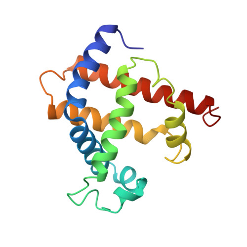
Solution NMR structures for sperm whale carbonmonoxy myoglobin have been calculated using 1301 distance restraints determined from nuclear Overhauser enhancement (NOE) measurements on 15N-labeled protein and chemical shift calculations for 385 protons. Starting structures included four crystal forms of myoglobin and 12 structures generated by metric matrix distance geometry. Refinements were also carried out using distance restraints alone. In general, the solution conformations are very close to the crystal structures, although the crystal structures are not consistent with some of the observed NOE connectivities. The solution structures are about as far apart from each other (as measured by backbone root-mean-square deviations) as they are from the crystal conformation. Inclusion of chemical shift restraints both tightened the spread of computed structures (especially in the heme pocket region) and led to structures that were closer to the X-ray conformation. The disposition of the side-chains near the heme group could in many cases be determined with considerable confidence, suggesting that a chemical shift analysis may be a useful adjunct to other sources of structural information available from NMR. In particular, this evidence suggests that the distal histidine residue is slightly displaced from the crystal conformation, but still inside the heme pocket at pH 5.6, that the side-chain of Leu89 is in contact with the heme ring but is probably disordered, and that the heme pocket where ligands bind is virtually identical in solution and in the crystal forms.
- Department of Molecular Biology, Scripps Research Institute, La Jolla, CA 92037.
Organizational Affiliation: