1MVF | pdb_00001mvf

MazE addiction antidote


Experimental Data Snapshot

  • Method: X-RAY DIFFRACTION
  • Resolution: 1.65 Å
  • R-Value Free: 
    0.249 (Depositor), 0.240 (DCC) 
  • R-Value Work: 
    0.218 (Depositor), 0.210 (DCC) 
  • R-Value Observed: 
    0.220 (Depositor) 

wwPDB Validation 3D Report Full Report

Validation slider image for 1MVF

This is version 1.3 of the entry. See complete history

Literature

Crystal structure of the intrinsically flexible addiction antidote MazE.

Loris, R.Marianovsky, I.Lah, J.Laeremans, T.Engelberg-Kulka, H.Glaser, G.Muyldermans, S.Wyns, L.

(2003) J Biological Chem 278: 28252-28257

  • DOI: https://doi.org/10.1074/jbc.M302336200
  • Primary Citation Related Structures: 
    1MVF

  • PubMed Abstract: 

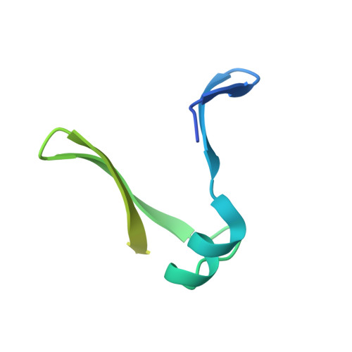
    A specific camel VHH (variable domain of dromedary heavy chain antibody) fragment was used to crystallize the intrinsically flexible addiction antidote MazE. Only 45% of the polypeptide chain is found ordered in the crystal. The MazE monomer consisting of two beta-hairpins connected by a short alpha-helix has no hydrophobic core on its own and represents only one half of a typical protein domain. A complete domain structure is formed by the association of two chains, creating a hydrophobic core between two four-stranded beta-sheets. This hydrophobic core consists exclusively of short aliphatic residues. The folded part of MazE contains a novel DNA binding motif. A model for DNA binding that is consistent with the available biochemical data is presented.


  • Organizational Affiliation
    • Laboratorium voor Ultrastructuur en Vlaams instituut voor Biotechnologie, Vrije Universiteit Brussel, Gebouw E, Pleinlaan 2, 1050 Brussel, Belgium. reloris@vub.ac.be

Macromolecule Content 

  • Total Structure Weight: 48.52 kDa 
  • Atom Count: 2,678 
  • Modeled Residue Count: 337 
  • Deposited Residue Count: 434 
  • Unique protein chains: 2

Macromolecules

Find similar proteins by:|  3D Structure
Entity ID: 1
MoleculeChains  Sequence LengthOrganismDetailsImage
immunoglobulin heavy chain variable region
A, B
135Camelus dromedariusMutation(s): 0 
Find similar proteins by:|  3D Structure
Entity ID: 2
MoleculeChains  Sequence LengthOrganismDetailsImage
PemI-like protein 1C [auth D],
D [auth E]
82Escherichia coliMutation(s): 0 
Gene Names: MazE
UniProt
Find proteins for P0AE72 (Escherichia coli (strain K12))
Explore P0AE72 
Go to UniProtKB:  P0AE72
Entity Groups
Sequence Clusters30% Identity50% Identity70% Identity90% Identity95% Identity100% Identity
UniProt GroupP0AE72
Sequence Annotations
Expand
Reference Sequence

Experimental Data & Validation

Experimental Data

  • Method: X-RAY DIFFRACTION
  • Resolution: 1.65 Å
  • R-Value Free:  0.249 (Depositor), 0.240 (DCC) 
  • R-Value Work:  0.218 (Depositor), 0.210 (DCC) 
  • R-Value Observed: 0.220 (Depositor) 
Space Group: P 1 21 1
Unit Cell:
Length ( Å )Angle ( ˚ )
a = 29.44α = 90
b = 47.826β = 90.591
c = 128.376γ = 90
Software Package:
Software NamePurpose
DENZOdata reduction
SCALEPACKdata scaling
AMoREphasing
CNSrefinement

Structure Validation

View Full Validation Report


Entry History 

Deposition Data

Revision History  (Full details and data files)

  • Version 1.0: 2003-06-10
    Type: Initial release
  • Version 1.1: 2008-04-28
    Changes: Version format compliance
  • Version 1.2: 2011-07-13
    Changes: Version format compliance
  • Version 1.3: 2024-10-30
    Changes: Data collection, Database references, Structure summary