1MPX | pdb_00001mpx

ALPHA-AMINO ACID ESTER HYDROLASE LABELED WITH SELENOMETHIONINE


Experimental Data Snapshot

  • Method: X-RAY DIFFRACTION
  • Resolution: 1.90 Å
  • R-Value Free: 
    0.178 (Depositor), 0.188 (DCC) 
  • R-Value Work: 
    0.149 (Depositor), 0.160 (DCC) 
  • R-Value Observed: 
    0.150 (Depositor) 

wwPDB Validation 3D Report Full Report

Validation slider image for 1MPX

This is version 1.4 of the entry. See complete history

Literature

The sequence and crystal structure of the alpha-amino acid ester hydrolase from Xanthomonas citri define a new family of beta-lactam antibiotic acylases.

Barends, T.R.Polderman-Tijmes, J.J.Jekel, P.A.Hensgens, C.M.de Vries, E.J.Janssen, D.B.Dijkstra, B.W.

(2003) J Biological Chem 278: 23076-23084

  • DOI: https://doi.org/10.1074/jbc.M302246200
  • Primary Citation Related Structures: 
    1MPX

  • PubMed Abstract: 

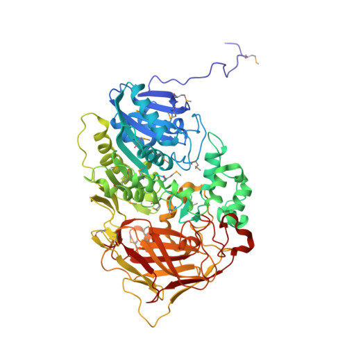
    alpha-Amino acid ester hydrolases (AEHs) catalyze the hydrolysis and synthesis of esters and amides with an alpha-amino group. As such, they can synthesize beta-lactam antibiotics from acyl compounds and beta-lactam nuclei obtained from the hydrolysis of natural antibiotics. This article describes the gene sequence and the 1.9-A resolution crystal structure of the AEH from Xanthomonas citri. The enzyme consists of an alpha/beta-hydrolase fold domain, a helical cap domain, and a jellyroll beta-domain. Structural homology was observed to the Rhodococcus cocaine esterase, indicating that both enzymes belong to the same class of bacterial hydrolases. Docking of a beta-lactam antibiotic in the active site explains the substrate specificity, specifically the necessity of an alpha-amino group on the substrate, and explains the low specificity toward the beta-lactam nucleus.


  • Organizational Affiliation
    • Department of Biophysical Chemistry, Groningen Biomolecular Sciences and Biotechnology Institute, University of Groningen, Nijenborgh 4, NL-9747 AG Groningen, The Netherlands.

Macromolecule Content 

  • Total Structure Weight: 279.6 kDa 
  • Atom Count: 21,702 
  • Modeled Residue Count: 2,456 
  • Deposited Residue Count: 2,460 
  • Unique protein chains: 1

Macromolecules

Find similar proteins by:|  3D Structure
Entity ID: 1
MoleculeChains  Sequence LengthOrganismDetailsImage
alpha-amino acid ester hydrolase
A, B, C, D
615Xanthomonas citriMutation(s): 18 
EC: 3.1.1.43
UniProt
Find proteins for Q6YBS3 (Xanthomonas citri)
Explore Q6YBS3 
Go to UniProtKB:  Q6YBS3
Entity Groups
Sequence Clusters30% Identity50% Identity70% Identity90% Identity95% Identity100% Identity
UniProt GroupQ6YBS3
Sequence Annotations
Expand
Reference Sequence

Small Molecules

Ligands 2 Unique
IDChains Name / Formula / InChI Key2D Diagram3D Interactions
GOL

Query on GOL


Download:Ideal Coordinates CCD File
F [auth A]
G [auth A]
H [auth A]
J [auth B]
K [auth B]
F [auth A],
G [auth A],
H [auth A],
J [auth B],
K [auth B],
L [auth B],
N [auth C],
O [auth C],
Q [auth D],
R [auth D]
GLYCEROL
C3 H8 O3
PEDCQBHIVMGVHV-UHFFFAOYSA-N
CA

Query on CA


Download:Ideal Coordinates CCD File
E [auth A],
I [auth B],
M [auth C],
P [auth D]
CALCIUM ION
Ca
BHPQYMZQTOCNFJ-UHFFFAOYSA-N
Modified Residues  1 Unique
IDChains TypeFormula2D DiagramParent
MSE
Query on MSE
A, B, C, D
L-PEPTIDE LINKINGC5 H11 N O2 SeMET

Experimental Data & Validation

Experimental Data

  • Method: X-RAY DIFFRACTION
  • Resolution: 1.90 Å
  • R-Value Free:  0.178 (Depositor), 0.188 (DCC) 
  • R-Value Work:  0.149 (Depositor), 0.160 (DCC) 
  • R-Value Observed: 0.150 (Depositor) 
Space Group: P 1 21 1
Unit Cell:
Length ( Å )Angle ( ˚ )
a = 89.772α = 90
b = 126.016β = 91.06
c = 132.291γ = 90
Software Package:
Software NamePurpose
DENZOdata reduction
SCALEPACKdata scaling
SnBphasing
SOLVEphasing
RESOLVEmodel building
ARP/wARPmodel building
REFMACrefinement
RESOLVEphasing

Structure Validation

View Full Validation Report


Entry History 

Deposition Data

Revision History  (Full details and data files)

  • Version 1.0: 2003-04-15
    Type: Initial release
  • Version 1.1: 2008-04-28
    Changes: Version format compliance
  • Version 1.2: 2011-07-13
    Changes: Non-polymer description, Version format compliance
  • Version 1.3: 2017-10-11
    Changes: Refinement description
  • Version 1.4: 2024-10-30
    Changes: Data collection, Database references, Derived calculations, Structure summary