1MOS | pdb_00001mos

ISOMERASE DOMAIN OF GLUCOSAMINE 6-PHOSPHATE SYNTHASE COMPLEXED WITH 2-AMINO-2-DEOXYGLUCITOL 6-PHOSPHATE


Experimental Data Snapshot

  • Method: X-RAY DIFFRACTION
  • Resolution: 2.00 Å
  • R-Value Free: 
    0.287 (Depositor) 
  • R-Value Observed: 
    0.244 (Depositor) 

Starting Model: experimental
View more details

wwPDB Validation 3D Report Full Report

Validation slider image for 1MOS

Ligand Structure Quality Assessment 


This is version 1.4 of the entry. See complete history

Literature

The mechanism of sugar phosphate isomerization by glucosamine 6-phosphate synthase.

Teplyakov, A.Obmolova, G.Badet-Denisot, M.A.Badet, B.

(1999) Protein Sci 8: 596-602

  • DOI: https://doi.org/10.1110/ps.8.3.596
  • Primary Citation Related Structures: 
    1MOR, 1MOS

  • PubMed Abstract: 

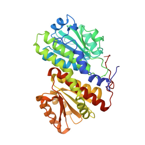
    Glucosamine 6-phosphate synthase converts fructose-6P into glucosamine-6P or glucose-6P depending on the presence or absence of glutamine. The isomerase activity is associated with a 40-kDa C-terminal domain, which has already been characterized crystallographically. Now the three-dimensional structures of the complexes with the reaction product glucose-6P and with the transition state analog 2-amino-2-deoxyglucitol-6P have been determined. Glucose-6P binds in a cyclic form whereas 2-amino-2-deoxyglucitol-6P is in an extended conformation. The information on ligand-protein interactions observed in the crystal structures together with the isotope exchange and site-directed mutagenesis data allow us to propose a mechanism of the isomerase activity of glucosamine-6P synthase. The sugar phosphate isomerization involves a ring opening step catalyzed by His504 and an enolization step with Glu488 catalyzing the hydrogen transfer from C1 to C2 of the substrate. The enediol intermediate is stabilized by a helix dipole and the epsilon-amino group of Lys603. Lys485 may play a role in deprotonating the hydroxyl O1 of the intermediate.


  • Organizational Affiliation
    • Genetics and Biochemistry Branch, National Institute of Diabetes and Digestive and Kidney Diseases, National Institutes of Health, Bethesda, Maryland 20892, USA. alext@gm-mv.niddk.nih.gov

Macromolecule Content 

  • Total Structure Weight: 41.12 kDa 
  • Atom Count: 3,048 
  • Modeled Residue Count: 367 
  • Deposited Residue Count: 368 
  • Unique protein chains: 1

Macromolecules

Find similar proteins by:|  3D Structure
Entity ID: 1
MoleculeChains  Sequence LengthOrganismDetailsImage
GLUCOSAMINE 6-PHOSPHATE SYNTHASE368Escherichia coliMutation(s): 0 
EC: 2.6.1.16
UniProt
Find proteins for P17169 (Escherichia coli (strain K12))
Explore P17169 
Go to UniProtKB:  P17169
Entity Groups
Sequence Clusters30% Identity50% Identity70% Identity90% Identity95% Identity100% Identity
UniProt GroupP17169
Sequence Annotations
Expand
Reference Sequence

Small Molecules

Ligands 4 Unique
IDChains Name / Formula / InChI Key2D Diagram3D Interactions
AGP

Query on AGP


Download:Ideal Coordinates CCD File
F [auth A]2-DEOXY-2-AMINO GLUCITOL-6-PHOSPHATE
C6 H16 N O8 P
LBNVXZROMBUNNQ-SLPGGIOYSA-N
MES

Query on MES


Download:Ideal Coordinates CCD File
G [auth A]2-(N-MORPHOLINO)-ETHANESULFONIC ACID
C6 H13 N O4 S
SXGZJKUKBWWHRA-UHFFFAOYSA-N
SO4

Query on SO4


Download:Ideal Coordinates CCD File
B [auth A],
C [auth A],
D [auth A]
SULFATE ION
O4 S
QAOWNCQODCNURD-UHFFFAOYSA-L
NA

Query on NA


Download:Ideal Coordinates CCD File
E [auth A]SODIUM ION
Na
FKNQFGJONOIPTF-UHFFFAOYSA-N

Experimental Data & Validation

Experimental Data

  • Method: X-RAY DIFFRACTION
  • Resolution: 2.00 Å
  • R-Value Free:  0.287 (Depositor) 
  • R-Value Observed: 0.244 (Depositor) 
Space Group: H 3 2
Unit Cell:
Length ( Å )Angle ( ˚ )
a = 143.9α = 90
b = 143.9β = 90
c = 172.8γ = 120
Software Package:
Software NamePurpose
CCP4model building
REFMACrefinement
DENZOdata reduction
SCALEPACKdata scaling
CCP4phasing

Structure Validation

View Full Validation Report


Ligand Structure Quality Assessment 


Entry History 

Deposition Data

Revision History  (Full details and data files)

  • Version 1.0: 1999-07-29
    Type: Initial release
  • Version 1.1: 2008-03-24
    Changes: Version format compliance
  • Version 1.2: 2011-07-13
    Changes: Derived calculations, Version format compliance
  • Version 1.3: 2023-08-09
    Changes: Data collection, Database references, Derived calculations, Other, Refinement description
  • Version 1.4: 2024-05-22
    Changes: Data collection