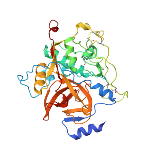
Structure of rat procathepsin B: model for inhibition of cysteine protease activity by the proregion.
Cygler, M., Sivaraman, J., Grochulski, P., Coulombe, R., Storer, A.C., Mort, J.S.(1996) Structure 4: 405-416
- PubMed: 8740363
- DOI: https://doi.org/10.1016/s0969-2126(96)00046-9
- Primary Citation of Related Structures:
1MIR - PubMed Abstract:
Cysteine proteases of the papain superfamily are synthesized as inactive precursors with a 60-110 residue N-terminal prosegment. The propeptides are potent inhibitors of their parent proteases. Although the proregion binding mode has been elucidated for all other protease classes, that of the cysteine proteases remained elusive. We report the three-dimensional structure of rat procathepsin B, determined at 2.8 A resolution. The 62-residue proregion does not form a globular structure on its own, but folds along the surface of mature cathepsin B. The N-terminal part of the proregion packs against a surface loop, with Trp24p (p indicating the proregion) playing a pivotal role in these interactions. Inhibition occurs by blocking access to the active site: part of the proregion enters the substrate-binding cleft in a similar manner to a natural substrate, but in a reverse orientation. The structure of procathepsin B provides the first insight into the mode of interaction between a mature cysteine protease from the papain superfamily and its prosegment. Maturation results in only one loop of cathepsin B changing conformation significantly, replacing contacts lost by removal of the prosegment. Contrary to many other proproteases, no rearrangement of the N terminus occurs following activation. Binding of the prosegment involves interaction with regions of the enzyme remote from the substrate-binding cleft and suggests a novel strategy for inhibitor design. The region of the prosegment where the activating cleavage occurs makes little contact with the enzyme, leading to speculation on the activation mechanism.
- Biotechnology Research Institute, National Research Council of Canada, Montreal, Quebec, Canada.
Organizational Affiliation:


