1MHH | pdb_00001mhh

Structure of P. magnus protein L mutant bound to a mouse Fab


Experimental Data Snapshot

  • Method: X-RAY DIFFRACTION
  • Resolution: 2.10 Å
  • R-Value Free: 
    0.247 (Depositor) 
  • R-Value Work: 
    0.197 (Depositor), 0.200 (DCC) 

wwPDB Validation 3D Report Full Report

Validation slider image for 1MHH

This is version 2.0 of the entry. See complete history

Literature

Evidence for plasticity and structural mimicry at the immunoglobulin light chain-protein L interface

Graille, M.Harrison, S.Crump, M.P.Findlow, S.C.Housden, N.G.Muller, B.H.Battail-Poirot, N.Sibai, G.Sutton, B.J.Taussig, M.J.Jolivet-Reynaud, C.Gore, M.G.Stura, E.A.

(2002) J Biological Chem 277: 47500-47506

  • DOI: https://doi.org/10.1074/jbc.M206105200
  • Primary Citation Related Structures: 
    1MHH

  • PubMed Abstract: 

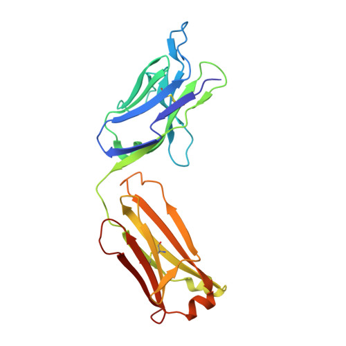
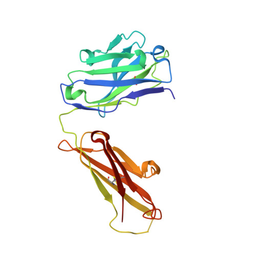
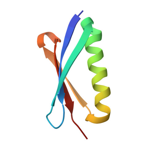
    The multidomain bacterial surface protein L (PpL) is a virulence factor expressed by only 10% of Peptostreptococcus magnus strains, and its expression is correlated with bacterial vaginosis. The molecular basis for its ability to recognize 60% of mammalian immunoglobulin light chain variable regions (V(L)) has been described recently by x-ray crystallography, which suggested the presence of two V(L) binding sites on each protein L domain (Graille, M., Stura, E. A., Housden, N. G., Beckingham, J. A., Bottomley, S. P., Beale, D., Taussig, M. J., Sutton, B. J., Gore, M. G., and Charbonnier, J. (2001) Structure 9, 679-687). Here, we report the crystal structure at 2.1 A resolution of a protein L mutant complexed to an Fab' fragment with only 50% of the V(L) residues interacting with PpL site 1 conserved. Comparison of the site 1 interface from both structures shows how protein L is able to accommodate these sequence differences and therefore bind to a large repertoire of Ig. The x-ray structure and NMR results confirm the existence of two V(L) binding sites on a single protein L domain. These sites exhibit a remarkable structural mimicry of growth factors binding to their receptors. This could explain the protein L superantigenic activity on human B lymphocytes.


  • Organizational Affiliation
    • Laboratoire de Structure des Protéines, Département d'Ingénierie et d'Etudes des Protéines (DIEP), Commissariat à l'Energie Atomique, Centre d'Etudes de Saclay, Gif-sur-Yvette, France.

Macromolecule Content 

  • Total Structure Weight: 109.55 kDa 
  • Atom Count: 8,490 
  • Modeled Residue Count: 994 
  • Deposited Residue Count: 1,000 
  • Unique protein chains: 3

Macromolecules

Find similar proteins by:|  3D Structure
Entity ID: 1
MoleculeChains  Sequence LengthOrganismDetailsImage
Fab, light chain
A, C
220Mus musculusMutation(s): 0 
Find similar proteins by:|  3D Structure
Entity ID: 2
MoleculeChains  Sequence LengthOrganismDetailsImage
Fab, heavy chain
B, D
217Mus musculusMutation(s): 0 
Find similar proteins by:|  3D Structure
Entity ID: 3
MoleculeChains  Sequence LengthOrganismDetailsImage
protein L domain C
E, F
63Finegoldia magna ATCC 29328Mutation(s): 0 

Small Molecules

Modified Residues  1 Unique
IDChains TypeFormula2D DiagramParent
AEA
Query on AEA
A, C
L-PEPTIDE LINKINGC5 H10 N2 O3 SCYS

Experimental Data & Validation

Experimental Data

  • Method: X-RAY DIFFRACTION
  • Resolution: 2.10 Å
  • R-Value Free:  0.247 (Depositor) 
  • R-Value Work:  0.197 (Depositor), 0.200 (DCC) 
Space Group: P 21 21 21
Unit Cell:
Length ( Å )Angle ( ˚ )
a = 78.486α = 90
b = 100.957β = 90
c = 149.147γ = 90
Software Package:
Software NamePurpose
DENZOdata reduction
SCALEPACKdata scaling
AMoREphasing
CNSrefinement

Structure Validation

View Full Validation Report


Entry History 

Deposition Data

Revision History  (Full details and data files)

  • Version 1.0: 2003-01-14
    Type: Initial release
  • Version 1.1: 2008-04-28
    Changes: Version format compliance
  • Version 1.2: 2011-07-13
    Changes: Source and taxonomy, Version format compliance
  • Version 1.3: 2018-01-03
    Changes: Database references, Structure summary
  • Version 2.0: 2025-03-26
    Changes: Data collection, Database references, Derived calculations, Polymer sequence, Structure summary