1MEM

Crystal structure of Cathepsin K complexed with a potent vinyl sulfone inhibitor


Experimental Data Snapshot

  • Method: X-RAY DIFFRACTION
  • Resolution: 1.80 Å
  • R-Value Work: 
    0.190 (DCC) 
  • R-Value Observed: 
    0.173 (Depositor) 

Starting Model: in silico
View more details

wwPDB Validation   3D Report Full Report


Ligand Structure Quality Assessment 

Created with Raphaël 2.3.0Worse 01 BetterLigand structure goodness of fit to experimental dataBest fitted 0D6Click on this verticalbar to view details

This is version 1.6 of the entry. See complete history


Literature

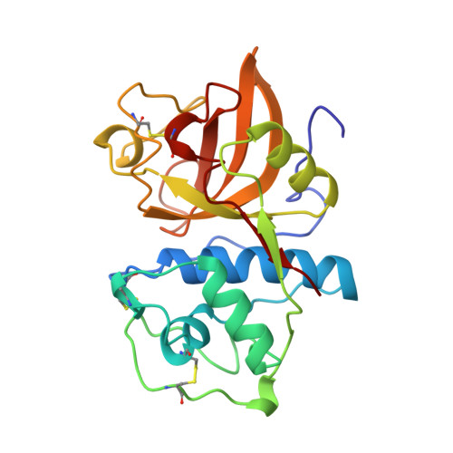
Crystal structure of human cathepsin K complexed with a potent inhibitor.

McGrath, M.E.Klaus, J.L.Barnes, M.G.Bromme, D.

(1997) Nat Struct Biol 4: 105-109


Macromolecules
Find similar proteins by:  (by identity cutoff)  |  3D Structure
Entity ID: 1
MoleculeChains Sequence LengthOrganismDetailsImage
Cathepsin K215Homo sapiensMutation(s): 0 
Gene Names: CTSKCTSOCTSO2
EC: 3.4.22.38
UniProt & NIH Common Fund Data Resources
Find proteins for P43235 (Homo sapiens)
Explore P43235 
Go to UniProtKB:  P43235
PHAROS:  P43235
GTEx:  ENSG00000143387 
Entity Groups  
Sequence Clusters30% Identity50% Identity70% Identity90% Identity95% Identity100% Identity
UniProt GroupP43235
Sequence Annotations
Expand
  • Reference Sequence
Small Molecules
Ligands 1 Unique
IDChains Name / Formula / InChI Key2D Diagram3D Interactions
0D6
Query on 0D6

Download Ideal Coordinates CCD File 
B [auth A]N-{(1R)-3-phenyl-1-[2-(phenylsulfonyl)ethyl]propyl}-N~2~-(piperazin-1-ylcarbonyl)-L-leucinamide
C28 H40 N4 O4 S
GZQYXFWTRHRNKT-AHWVRZQESA-N
Experimental Data & Validation

Experimental Data

  • Method: X-RAY DIFFRACTION
  • Resolution: 1.80 Å
  • R-Value Work:  0.190 (DCC) 
  • R-Value Observed: 0.173 (Depositor) 
Space Group: P 21 21 21
Unit Cell:
Length ( Å )Angle ( ˚ )
a = 40.82α = 90
b = 50.04β = 90
c = 105.92γ = 90
Software Package:
Software NamePurpose
bioteXdata collection
MSCdata collection
bioteXdata reduction
MSCdata reduction
X-PLORmodel building
X-PLORrefinement
bioteXdata scaling
X-PLORphasing

Structure Validation

View Full Validation Report


Ligand Structure Quality Assessment 

Created with Raphaël 2.3.0Worse 01 BetterLigand structure goodness of fit to experimental dataBest fitted 0D6Click on this verticalbar to view details

Entry History 

Deposition Data

Revision History  (Full details and data files)

  • Version 1.0: 1998-01-14
    Type: Initial release
  • Version 1.1: 2008-03-24
    Changes: Version format compliance
  • Version 1.2: 2011-07-13
    Changes: Atomic model, Database references, Derived calculations, Non-polymer description, Structure summary, Version format compliance
  • Version 1.3: 2012-12-12
    Changes: Other
  • Version 1.4: 2014-01-29
    Changes: Refinement description
  • Version 1.5: 2024-04-03
    Changes: Data collection, Database references, Derived calculations, Other, Refinement description
  • Version 1.6: 2024-11-06
    Changes: Structure summary