1MD9 | pdb_00001md9

CRYSTAL STRUCTURE OF DhbE IN COMPLEX WITH DHB AND AMP


Experimental Data Snapshot

  • Method: X-RAY DIFFRACTION
  • Resolution: 2.80 Å
  • R-Value Free: 
    0.288 (Depositor) 
  • R-Value Work: 
    0.232 (Depositor) 

wwPDB Validation 3D Report Full Report

Validation slider image for 1MD9

This is version 1.4 of the entry. See complete history

Literature

Crystal structure of DhbE, an archetype for aryl acid activating domains of modular nonribosomal peptide synthetases.

May, J.J.Kessler, N.Marahiel, M.A.Stubbs, M.T.

(2002) Proc Natl Acad Sci U S A 99: 12120-12125

  • DOI: https://doi.org/10.1073/pnas.182156699
  • Primary Citation Related Structures: 
    1MD9, 1MDB, 1MDF

  • PubMed Abstract: 

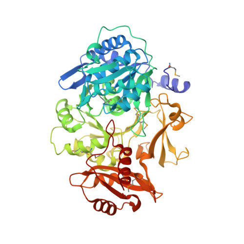
    The synthesis of the catecholic siderophore bacillibactin is accomplished by the nonribosomal peptide synthetase (NRPS) encoded by the dhb operon. DhbE is responsible for the initial step in bacillibactin synthesis, the activation of the aryl acid 2,3-dihydroxybenzoate (DHB). The stand-alone adenylation (A) domain DhbE, the structure of which is presented here, exhibits greatest homology to other NRPS A-domains, acyl-CoA ligases and luciferases. It's structure is solved in three different states, without the ligands ATP and DHB (native state), with the product DHB-AMP (adenylate state) and with the hydrolyzed product AMP and DHB (hydrolyzed state). The 59.9-kDa protein folds into two domains, with the active site at the interface between them. In contrast to previous proposals of a major reorientation of the large and small domains on substrate binding, we observe only local structural rearrangements. The structure of the phosphate binding loop could be determined, a motif common to many adenylate-forming enzymes, as well as with bound DHB-adenylate and the hydrolyzed product DHB*AMP. Based on the structure and amino acid sequence alignments, an adapted specificity conferring code for aryl acid activating domains is proposed, allowing assignment of substrate specificity to gene products of previously unknown function.


  • Organizational Affiliation
    • Biochemie, Fachbereich Chemie, Philipps-Universität Marburg, Hans-Meerwein-Strasse, 35032 Marburg, Germany.

Macromolecule Content 

  • Total Structure Weight: 60.86 kDa 
  • Atom Count: 4,232 
  • Modeled Residue Count: 536 
  • Deposited Residue Count: 539 
  • Unique protein chains: 1

Macromolecules

Find similar proteins by:|  3D Structure
Entity ID: 1
MoleculeChains  Sequence LengthOrganismDetailsImage
2,3-dihydroxybenzoate-AMP ligase539Bacillus subtilisMutation(s): 0 
EC: 6.3.2 (PDB Primary Data), 6.2.1.71 (UniProt)
UniProt
Find proteins for P40871 (Bacillus subtilis (strain 168))
Explore P40871 
Go to UniProtKB:  P40871
Entity Groups
Sequence Clusters30% Identity50% Identity70% Identity90% Identity95% Identity100% Identity
UniProt GroupP40871
Sequence Annotations
Expand
Reference Sequence

Small Molecules

Ligands 2 Unique
IDChains Name / Formula / InChI Key2D Diagram3D Interactions
AMP

Query on AMP


Download:Ideal Coordinates CCD File
B [auth A]ADENOSINE MONOPHOSPHATE
C10 H14 N5 O7 P
UDMBCSSLTHHNCD-KQYNXXCUSA-N
DBH

Query on DBH


Download:Ideal Coordinates CCD File
C [auth A]2,3-DIHYDROXY-BENZOIC ACID
C7 H6 O4
GLDQAMYCGOIJDV-UHFFFAOYSA-N
Modified Residues  1 Unique
IDChains TypeFormula2D DiagramParent
MSE
Query on MSE
A
L-PEPTIDE LINKINGC5 H11 N O2 SeMET

Experimental Data & Validation

Experimental Data

  • Method: X-RAY DIFFRACTION
  • Resolution: 2.80 Å
  • R-Value Free:  0.288 (Depositor) 
  • R-Value Work:  0.232 (Depositor) 
Space Group: P 31
Unit Cell:
Length ( Å )Angle ( ˚ )
a = 66.9α = 90
b = 66.9β = 90
c = 114.8γ = 120
Software Package:
Software NamePurpose
DENZOdata reduction
SCALEPACKdata scaling
CNSrefinement

Structure Validation

View Full Validation Report


Entry History 

Deposition Data

Revision History  (Full details and data files)

  • Version 1.0: 2002-09-11
    Type: Initial release
  • Version 1.1: 2008-04-28
    Changes: Version format compliance
  • Version 1.2: 2011-07-13
    Changes: Version format compliance
  • Version 1.3: 2017-10-11
    Changes: Refinement description
  • Version 1.4: 2024-12-25
    Changes: Advisory, Data collection, Database references, Derived calculations, Structure summary