1MCW

THREE-DIMENSIONAL STRUCTURE OF A HYBRID LIGHT CHAIN DIMER. PROTEIN ENGINEERING OF A BINDING CAVITY


Experimental Data Snapshot

  • Method: X-RAY DIFFRACTION
  • Resolution: 3.50 Å
  • R-Value Observed: 0.170 

wwPDB Validation   3D Report Full Report


This is version 2.0 of the entry. See complete history


Literature

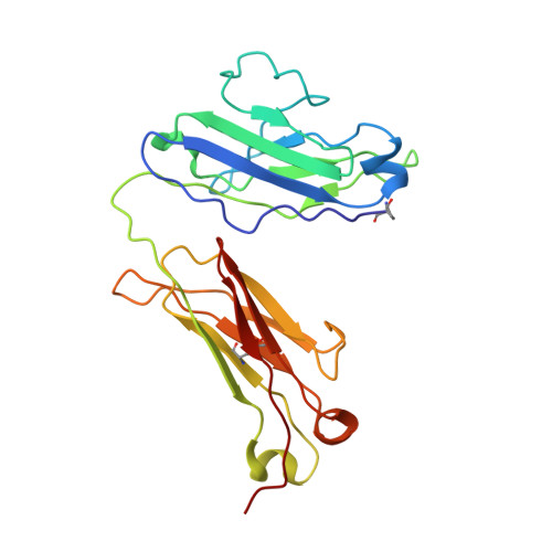
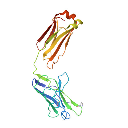
Three-dimensional structure of a hybrid light chain dimer: protein engineering of a binding cavity.

Ely, K.R.Herron, J.N.Edmundson, A.B.

(1990) Mol Immunol 27: 101-114

  • DOI: https://doi.org/10.1016/0161-5890(90)90105-9
  • Primary Citation of Related Structures:  
    1MCW

  • PubMed Abstract: 

    An attempt was made to engineer a binding site and check its structure by X-ray analysis. Two human light chains (Mcg and Weir), with "variable" domain sequences differing in 36 positions, were hybridized into a heterologous dimer and crystallized in ammonium sulfate by the same procedure used for the trigonal form of the Mcg dimer. The three-dimensional structure of the hybrid was determined at 3.5-A resolution by difference Fourier analysis, interactive model building with computer graphics and crystallographic refinement. In the heterologous dimer, the Weir protein behaved as the structural analog of the heavy chain in an antigen binding fragment, while the Mcg protein assumed the role of the light chain component. The hybrid and the Mcg dimer were closely similar in overall structure, an observation probably correlated with the deliberate cleavage of the intrachain disulfide bond in the variable domain of the Weir protein during the hybridization procedure. Examination of the crystal structure of the hybrid suggested that the cleavage resulted in the relaxation of restraints which might otherwise have interfered with the formation of an Mcg-like dimer. There were six substitutions among the residues lining the binding cavities of the hybrid and Mcg dimer. These substitutions significantly affected the sizes, shapes and binding properties of the two cavities.


  • Organizational Affiliation

    Department of Biology, University of Utah, Salt Lake City 84112.


Macromolecules
Find similar proteins by:  (by identity cutoff)  |  3D Structure
Entity ID: 1
MoleculeChains Sequence LengthOrganismDetailsImage
IMMUNOGLOBULIN WEIR (LIGHT CHAIN)A [auth W]216Homo sapiensMutation(s): 0 
Entity Groups  
Sequence Clusters30% Identity50% Identity70% Identity90% Identity95% Identity100% Identity
Sequence Annotations
Expand
  • Reference Sequence
Find similar proteins by:  (by identity cutoff)  |  3D Structure
Entity ID: 2
MoleculeChains Sequence LengthOrganismDetailsImage
IMMUNOGLOBULIN MCG (LIGHT CHAIN)B [auth M]216Homo sapiensMutation(s): 0 
UniProt & NIH Common Fund Data Resources
Find proteins for P01709 (Homo sapiens)
Explore P01709 
Go to UniProtKB:  P01709
GTEx:  ENSG00000278196 
Entity Groups  
Sequence Clusters30% Identity50% Identity70% Identity90% Identity95% Identity100% Identity
UniProt GroupP01709
Sequence Annotations
Expand
  • Reference Sequence
Small Molecules
Modified Residues  1 Unique
IDChains TypeFormula2D DiagramParent
PCA
Query on PCA
A [auth W]L-PEPTIDE LINKINGC5 H7 N O3GLN
Experimental Data & Validation

Experimental Data

  • Method: X-RAY DIFFRACTION
  • Resolution: 3.50 Å
  • R-Value Observed: 0.170 
  • Space Group: P 31 2 1
Unit Cell:
Length ( Å )Angle ( ˚ )
a = 72.3α = 90
b = 72.3β = 90
c = 185.9γ = 120
Software Package:
Software NamePurpose
PROLSQrefinement

Structure Validation

View Full Validation Report


Entry History 

Deposition Data

Revision History  (Full details and data files)

  • Version 1.0: 1990-10-15
    Type: Initial release
  • Version 1.1: 2008-03-24
    Changes: Version format compliance
  • Version 1.2: 2011-07-13
    Changes: Version format compliance
  • Version 2.0: 2019-12-25
    Changes: Database references, Derived calculations, Other, Polymer sequence