1M0W | pdb_00001m0w

Yeast Glutathione Synthase Bound to gamma-glutamyl-cysteine, AMP-PNP and 2 Magnesium Ions


Experimental Data Snapshot

  • Method: X-RAY DIFFRACTION
  • Resolution: 1.80 Å
  • R-Value Free: 
    0.196 (Depositor) 
  • R-Value Work: 
    0.172 (Depositor) 
  • R-Value Observed: 
    0.172 (Depositor) 

Starting Model: experimental
View more details

wwPDB Validation 3D Report Full Report

Validation slider image for 1M0W

This is version 1.4 of the entry. See complete history

Literature

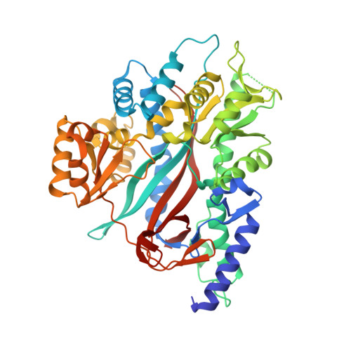
Large Conformational Changes in the Catalytic Cycle of Glutathione Synthase

Gogos, A.Shapiro, L.

(2002) Structure 10: 1669-1676

  • DOI: https://doi.org/10.1016/s0969-2126(02)00906-1
  • Primary Citation Related Structures: 
    1M0T, 1M0W

  • PubMed Abstract: 

    Glutathione synthase catalyzes the final ATP-dependent step in glutathione biosynthesis, the formation of glutathione from gamma-glutamylcysteine and glycine. We have determined structures of yeast glutathione synthase in two forms: unbound (2.3 A resolution) and bound to its substrate gamma-glutamylcysteine, the ATP analog AMP-PNP, and two magnesium ions (1.8 A resolution). These structures reveal that upon substrate binding, large domain motions convert the enzyme from an open unliganded form to a closed conformation in which protein domains completely surround the substrate in the active site.


  • Organizational Affiliation
    • Department of Biochemistry and Molecular Biophysics, Columbia University College of Physicians and Surgeons, 630 West 168th Street, New York, NY 10032, USA.

Macromolecule Content 

  • Total Structure Weight: 114.04 kDa 
  • Atom Count: 8,810 
  • Modeled Residue Count: 960 
  • Deposited Residue Count: 982 
  • Unique protein chains: 1

Macromolecules

Find similar proteins by:|  3D Structure
Entity ID: 1
MoleculeChains  Sequence LengthOrganismDetailsImage
glutathione synthetase
A, B
491Saccharomyces cerevisiaeMutation(s): 0 
Gene Names: GSH2
EC: 6.3.2.3
UniProt
Find proteins for Q08220 (Saccharomyces cerevisiae (strain ATCC 204508 / S288c))
Explore Q08220 
Go to UniProtKB:  Q08220
Entity Groups
Sequence Clusters30% Identity50% Identity70% Identity90% Identity95% Identity100% Identity
UniProt GroupQ08220
Sequence Annotations
Expand
Reference Sequence

Small Molecules

Ligands 4 Unique
IDChains Name / Formula / InChI Key2D Diagram3D Interactions
ANP

Query on ANP


Download:Ideal Coordinates CCD File
H [auth A],
P [auth B]
PHOSPHOAMINOPHOSPHONIC ACID-ADENYLATE ESTER
C10 H17 N6 O12 P3
PVKSNHVPLWYQGJ-KQYNXXCUSA-N
3GC

Query on 3GC


Download:Ideal Coordinates CCD File
I [auth A],
Q [auth B]
GAMMA-GLUTAMYLCYSTEINE
C8 H14 N2 O5 S
RITKHVBHSGLULN-WHFBIAKZSA-N
SO4

Query on SO4


Download:Ideal Coordinates CCD File
E [auth A]
F [auth A]
G [auth A]
L [auth B]
M [auth B]
E [auth A],
F [auth A],
G [auth A],
L [auth B],
M [auth B],
N [auth B],
O [auth B]
SULFATE ION
O4 S
QAOWNCQODCNURD-UHFFFAOYSA-L
MG

Query on MG


Download:Ideal Coordinates CCD File
C [auth A],
D [auth A],
J [auth B],
K [auth B]
MAGNESIUM ION
Mg
JLVVSXFLKOJNIY-UHFFFAOYSA-N

Experimental Data & Validation

Experimental Data

  • Method: X-RAY DIFFRACTION
  • Resolution: 1.80 Å
  • R-Value Free:  0.196 (Depositor) 
  • R-Value Work:  0.172 (Depositor) 
  • R-Value Observed: 0.172 (Depositor) 
Space Group: P 1
Unit Cell:
Length ( Å )Angle ( ˚ )
a = 51.661α = 82.08
b = 52.006β = 86.77
c = 100.636γ = 77.49
Software Package:
Software NamePurpose
DENZOdata reduction
SCALEPACKdata scaling
AMoREphasing
CNSrefinement

Structure Validation

View Full Validation Report


Entry History 

Deposition Data

Revision History  (Full details and data files)

  • Version 1.0: 2002-12-11
    Type: Initial release
  • Version 1.1: 2008-04-28
    Changes: Version format compliance
  • Version 1.2: 2011-07-13
    Changes: Version format compliance
  • Version 1.3: 2021-02-03
    Changes: Derived calculations, Structure summary
  • Version 1.4: 2024-02-14
    Changes: Data collection, Database references, Refinement description