1M0V | pdb_00001m0v

NMR STRUCTURE OF THE TYPE III SECRETORY DOMAIN OF YERSINIA YOPH COMPLEXED WITH THE SKAP-HOM PHOSPHO-PEPTIDE N-acetyl-DEpYDDPF-NH2


Experimental Data Snapshot

  • Method: SOLUTION NMR
  • Conformers Calculated: 360 
  • Conformers Submitted: 20 
  • Selection Criteria: structures with the least restraint violations 

wwPDB Validation 3D Report Full Report

Validation slider image for 1M0V

This is version 1.4 of the entry. See complete history

Literature

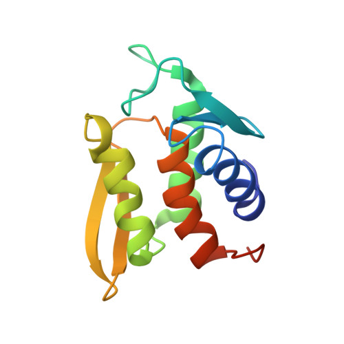
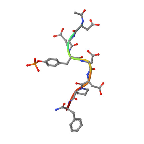
Solution structure and phosphopeptide binding to the N-terminal domain of Yersinia YopH: comparison with a crystal structure

Khandelwal, P.Keliikuli, K.Smith, C.L.Saper, M.A.Zuiderweg, E.R.P.

(2002) Biochemistry 41: 11425-11437

  • DOI: https://doi.org/10.1021/bi026333l
  • Primary Citation Related Structures: 
    1M0V

  • PubMed Abstract: 

    Virulence of pathogenic bacteria of the genus Yersinia requires the injection of six effector proteins into the cytoplasm of host cells. The amino-terminal domain of one of these effectors, the tyrosine phosphatase YopH, is essential for translocation of YopH, as well as for targeting it to phosphotyrosine-containing substrates of the type pYxxP. We report the high-resolution solution structure of the N-terminal domain (residues 1-129) from the Yersinia pseudotuberculosis YopH (YopH-NT) in complex with N-acetyl-DEpYDDPF-NH(2), a peptide derived from an in vivo protein substrate. In contrast to the domain-swapped dimer observed in a crystal structure of the same protein (Smith, C. L., Khandelwal, P., Keliikuli, K., Zuiderweg, E. R. P., and Saper, M. A. (2001) Mol. Microbiol. 42, 967-979), YopH-NT is monomeric in solution. The peptide binding site is located on a beta-hairpin that becomes the crossover point in the dimer structure. The binding site has several characteristics that are reminiscent of SH2 domains, which also bind to pYxxP sequences.


  • Organizational Affiliation
    • Biophysics Research Division, University of Michigan, 930 North University Avenue, Ann Arbor, Michigan 48109-1055, USA.

Macromolecule Content 

  • Total Structure Weight: 15.89 kDa 
  • Atom Count: 1,053 
  • Modeled Residue Count: 139 
  • Deposited Residue Count: 145 
  • Unique protein chains: 2

Macromolecules

Find similar proteins by:|  3D Structure
Entity ID: 1
MoleculeChains  Sequence LengthOrganismDetailsImage
PROTEIN-TYROSINE PHOSPHATASE YOPH136Yersinia pseudotuberculosisMutation(s): 0 
EC: 3.1.3.48
UniProt
Find proteins for P08538 (Yersinia pseudotuberculosis serotype I (strain IP32953))
Explore P08538 
Go to UniProtKB:  P08538
Entity Groups
Sequence Clusters30% Identity50% Identity70% Identity90% Identity95% Identity100% Identity
UniProt GroupP08538
Sequence Annotations
Expand
Reference Sequence
Find similar proteins by:  Sequence   |   3D Structure  
Entity ID: 2
MoleculeChains  Sequence LengthOrganismDetailsImage
SKAP55 homologue9N/AMutation(s): 1 
UniProt
Find proteins for Q3UND0 (Mus musculus)
Explore Q3UND0 
Go to UniProtKB:  Q3UND0
Entity Groups
UniProt GroupQ3UND0
Sequence Annotations
Expand
Reference Sequence

Small Molecules

Modified Residues  1 Unique
IDChains TypeFormula2D DiagramParent
PTR
Query on PTR
B
L-PEPTIDE LINKINGC9 H12 N O6 PTYR

Experimental Data & Validation

Experimental Data

  • Method: SOLUTION NMR
  • Conformers Calculated: 360 
  • Conformers Submitted: 20 
  • Selection Criteria: structures with the least restraint violations 

Structure Validation

View Full Validation Report


Entry History 

Deposition Data

Revision History  (Full details and data files)

  • Version 1.0: 2002-07-24
    Type: Initial release
  • Version 1.1: 2008-04-28
    Changes: Version format compliance
  • Version 1.2: 2011-07-13
    Changes: Version format compliance
  • Version 1.3: 2022-02-23
    Changes: Data collection, Database references, Derived calculations
  • Version 1.4: 2024-11-20
    Changes: Data collection, Structure summary