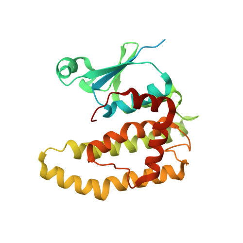
Structure of a Drosophila Sigma Class Glutathione S-transferase Reveals a Novel Active Site Topography Suited for Lipid Peroxidation Products
Agianian, B., Tucker, P.A., Schouten, A., Leonard, K., Bullard, B., Gros, P.(2003) J Mol Biology 326: 151-165
- PubMed: 12547198 Search on PubMed
- DOI: https://doi.org/10.1016/s0022-2836(02)01327-x
- Primary Citation Related Structures:
1M0U - PubMed Abstract:
Insect glutathione-S-transferases (GSTs) are grouped in three classes, I, II and recently III; class I (Delta class) enzymes together with class III members are implicated in conferring resistance to insecticides. Class II (Sigma class) GSTs, however, are poorly characterized and their exact biological function remains elusive. Drosophila glutathione S-transferase-2 (GST-2) (DmGSTS1-1) is a class II enzyme previously found associated specifically with the insect indirect flight muscle. It was recently shown that GST-2 exhibits considerable conjugation activity for 4-hydroxynonenal (4-HNE), a lipid peroxidation product, raising the possibility that it has a major anti-oxidant role in the flight muscle. Here, we report the crystal structure of GST-2 at 1.75A resolution. The GST-2 dimer shows the canonical GST fold with glutathione (GSH) ordered in only one of the two binding sites. While the GSH-binding mode is similar to other GST structures, a distinct orientation of helix alpha6 creates a novel electrophilic substrate-binding site (H-site) topography, largely flat and without a prominent hydrophobic-binding pocket, which characterizes the H-sites of other GSTs. The H-site displays directionality in the distribution of charged/polar and hydrophobic residues creating a binding surface that explains the selectivity for amphipolar peroxidation products, with the polar-binding region formed by residues Y208, Y153 and R145 and the hydrophobic-binding region by residues V57, A59, Y211 and the C-terminal V249. A structure-based model of 4-HNE binding is presented. The model suggest that residues Y208, R145 and possibly Y153 may be key residues involved in catalysis.
- Department of Crystal and Structural Chemistry, Bijvoet Center for Biomolecular Research, Utrecht University, Padualaan 8, 3584 CH, Utrecht, The Netherlands. agianian@embl-heidelberg.de
Organizational Affiliation: