1LZN | pdb_00001lzn

NEUTRON STRUCTURE OF HEN EGG-WHITE LYSOZYME


Experimental Data Snapshot

  • Method: NEUTRON DIFFRACTION
  • Resolution: 1.70 Å
  • R-Value Free: 
    0.221 (Depositor) 
  • R-Value Work: 
    0.204 (Depositor) 

Starting Model: experimental
View more details

wwPDB Validation 3D Report Full Report

Validation slider image for 1LZN

This is version 1.6 of the entry. See complete history

Literature

Quasi-Laue neutron-diffraction study of the water arrangement in crystals of triclinic hen egg-white lysozyme.

Bon, C.Lehmann, M.S.Wilkinson, C.

(1999) Acta Crystallogr D Biol Crystallogr 55: 978-987

  • DOI: https://doi.org/10.1107/s0907444998018514
  • Primary Citation Related Structures: 
    1LZN

  • PubMed Abstract: 

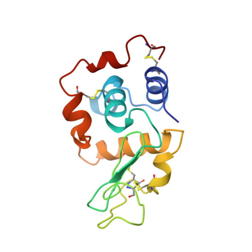
    Triclinic crystals of lysozyme, hydrogen-deuterium exchanged in deuterated solvent, have been studied using neutron quasi-Laue techniques and a newly developed cylinder image-plate detector. The wavelength range employed was from 2.7 to 3.5 A, which gave 9426 significant reflections [F >/= 2sigma(F)] to a resolution limit of 1. 7 A. The deuteration states of the H atoms in the protein molecule were identified, followed by an extensive analysis of the water structure surrounding the protein. The final R factor was 20.4% (Rfree = 22.1%). In total, the 244 observed water molecules form approximately one layer of water around the protein with far fewer water molecules located further away. Water molecules covering the apolar patches make tangential layers at 4-5 A from the surface or form C-H...O contacts, and several water-molecule sites can be identified in the apolar cavities. Many of the water molecules are apparently orientationally disordered, and only 115 out of the 244 water molecules sit in mean single orientations. Comparison of these results with quasi-elastic neutron scattering observations of the water dynamics leads to a picture of the water molecules forming an extended constantly fluctuating network covering the protein surface.


  • Organizational Affiliation
    • Université Joseph Fourier, F-38400 Saint Martin d'Hères, France and Institut Laue-Langevin, F-38042 Grenoble, France.

Macromolecule Content 

  • Total Structure Weight: 14.66 kDa 
  • Atom Count: 1,265 
  • Modeled Residue Count: 129 
  • Deposited Residue Count: 129 
  • Unique protein chains: 1

Macromolecules

Find similar proteins by:|  3D Structure
Entity ID: 1
MoleculeChains  Sequence LengthOrganismDetailsImage
PROTEIN (LYSOZYME)129Gallus gallusMutation(s): 0 
EC: 3.2.1.17
UniProt
Find proteins for P00698 (Gallus gallus)
Explore P00698 
Go to UniProtKB:  P00698
Entity Groups
Sequence Clusters30% Identity50% Identity70% Identity90% Identity95% Identity100% Identity
UniProt GroupP00698
Sequence Annotations
Expand
Reference Sequence

Experimental Data & Validation

Experimental Data

  • Method: NEUTRON DIFFRACTION
  • Resolution: 1.70 Å
  • R-Value Free:  0.221 (Depositor) 
  • R-Value Work:  0.204 (Depositor) 
Space Group: P 1
Unit Cell:
Length ( Å )Angle ( ˚ )
a = 27.28α = 88.8
b = 32.04β = 108.8
c = 34.27γ = 111.6
Software Package:
Software NamePurpose
CCP4refinement
CCP4data reduction
X-PLORrefinement
CCP4data scaling

Structure Validation

View Full Validation Report


Entry History 

Deposition Data

Revision History  (Full details and data files)

  • Version 1.0: 1999-04-01
    Type: Initial release
  • Version 1.1: 2008-04-26
    Changes: Version format compliance
  • Version 1.2: 2011-07-13
    Changes: Non-polymer description, Version format compliance
  • Version 1.3: 2017-10-04
    Changes: Refinement description
  • Version 1.4: 2021-06-30
    Changes: Data collection, Derived calculations
  • Version 1.5: 2023-12-27
    Changes: Data collection, Database references, Refinement description
  • Version 1.6: 2024-11-13
    Changes: Structure summary