1LT8 | pdb_00001lt8

Reduced Homo sapiens Betaine-Homocysteine S-Methyltransferase in Complex with S-(delta-carboxybutyl)-L-Homocysteine


Experimental Data Snapshot

  • Method: X-RAY DIFFRACTION
  • Resolution: 2.05 Å
  • R-Value Free: 
    0.255 (Depositor), 0.251 (DCC) 
  • R-Value Work: 
    0.226 (Depositor), 0.225 (DCC) 
  • R-Value Observed: 
    0.236 (Depositor) 

Starting Model: experimental
View more details

wwPDB Validation 3D Report Full Report

Validation slider image for 1LT8

Ligand Structure Quality Assessment 


This is version 1.4 of the entry. See complete history

Literature

Betaine-homocysteine methyltransferase: zinc in a distorted barrel.

Evans, J.C.Huddler, D.P.Jiracek, J.Castro, C.Millian, N.S.Garrow, T.A.Ludwig, M.L.

(2002) Structure 10: 1159-1171

  • DOI: https://doi.org/10.1016/s0969-2126(02)00796-7
  • Primary Citation Related Structures: 
    1LT7, 1LT8

  • PubMed Abstract: 

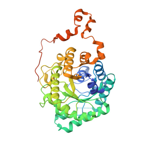
    Betaine-homocysteine methyl transferase (BHMT) catalyzes the synthesis of methionine from betaine and homocysteine (Hcy), utilizing a zinc ion to activate Hcy. BHMT is a key liver enzyme that is important for homocysteine homeostasis. X-ray structures of human BHMT in its oxidized (Zn-free) and reduced (Zn-replete) forms, the latter in complex with the bisubstrate analog, S(delta-carboxybutyl)-L-homocysteine, were determined at resolutions of 2.15 A and 2.05 A. BHMT is a (beta/alpha)(8) barrel that is distorted to construct the substrate and metal binding sites. The zinc binding sequences G-V/L-N-C and G-G-C-C are at the C termini of strands beta6 and beta8. Oxidation to the Cys217-Cys299 disulfide and expulsion of Zn are accompanied by local rearrangements. The structures identify Hcy binding fingerprints and provide a prototype for the homocysteine S-methyltransferase family.


  • Organizational Affiliation
    • Biophysics Research Division and Department of Biological Chemistry, University of Michigan, Ann Arbor, MI 48109, USA.

Macromolecule Content 

  • Total Structure Weight: 90.67 kDa 
  • Atom Count: 5,826 
  • Modeled Residue Count: 688 
  • Deposited Residue Count: 812 
  • Unique protein chains: 1

Macromolecules

Find similar proteins by:|  3D Structure
Entity ID: 1
MoleculeChains  Sequence LengthOrganismDetailsImage
BETAINE-HOMOCYSTEINE METHYLTRANSFERASE
A, B
406Homo sapiensMutation(s): 6 
EC: 2.1.1.5
UniProt & NIH Common Fund Data Resources
Find proteins for Q93088 (Homo sapiens)
Explore Q93088 
Go to UniProtKB:  Q93088
PHAROS:  Q93088
GTEx:  ENSG00000145692 
Entity Groups
Sequence Clusters30% Identity50% Identity70% Identity90% Identity95% Identity100% Identity
UniProt GroupQ93088
Sequence Annotations
Expand
Reference Sequence

Small Molecules

Binding Affinity Annotations 
IDSourceBinding Affinity
CBH BindingDB:  1LT8 Ki: 12 (nM) from 1 assay(s)
Kd: 280 (nM) from 1 assay(s)
IC50: min: 87, max: 138 (nM) from 3 assay(s)

Experimental Data & Validation

Experimental Data

  • Method: X-RAY DIFFRACTION
  • Resolution: 2.05 Å
  • R-Value Free:  0.255 (Depositor), 0.251 (DCC) 
  • R-Value Work:  0.226 (Depositor), 0.225 (DCC) 
  • R-Value Observed: 0.236 (Depositor) 
Space Group: C 1 2 1
Unit Cell:
Length ( Å )Angle ( ˚ )
a = 109.702α = 90
b = 90.836β = 122.28
c = 88.403γ = 90
Software Package:
Software NamePurpose
CNSrefinement
DENZOdata reduction
SCALEPACKdata scaling
CNSphasing

Structure Validation

View Full Validation Report


Ligand Structure Quality Assessment 


Entry History 

Deposition Data

Revision History  (Full details and data files)

  • Version 1.0: 2002-09-11
    Type: Initial release
  • Version 1.1: 2008-04-28
    Changes: Version format compliance
  • Version 1.2: 2011-07-13
    Changes: Version format compliance
  • Version 1.3: 2021-10-27
    Changes: Database references, Derived calculations
  • Version 1.4: 2024-02-14
    Changes: Data collection, Refinement description