1LQA | pdb_00001lqa

TAS PROTEIN FROM ESCHERICHIA COLI IN COMPLEX WITH NADPH


Experimental Data Snapshot

  • Method: X-RAY DIFFRACTION
  • Resolution: 1.60 Å
  • R-Value Free: 
    0.186 (Depositor), 0.190 (DCC) 
  • R-Value Work: 
    0.145 (Depositor), 0.150 (DCC) 
  • R-Value Observed: 
    0.146 (Depositor) 

wwPDB Validation 3D Report Full Report

Validation slider image for 1LQA

Ligand Structure Quality Assessment 


This is version 1.3 of the entry. See complete history

Literature

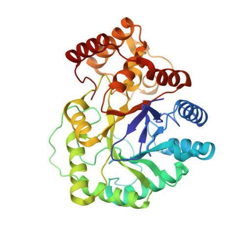
Crystal structure of the Escherichia coli Tas protein, an NADP(H)-dependent aldo-keto reductase

Obmolova, G.Teplyakov, A.Khil, P.P.Howard, A.J.Camerini-Otero, R.D.Gilliland, G.L.

(2003) Proteins 53: 323-325

  • DOI: https://doi.org/10.1186/1472-6807-3-7
  • Primary Citation Related Structures: 
    1LQA, 1NMO, 1NMP

  • PubMed Abstract: 

    The protein encoded by the gene ybgI was chosen as a target for a structural genomics project emphasizing the relation of protein structure to function. The structure of the ybgI protein is a toroid composed of six polypeptide chains forming a trimer of dimers. Each polypeptide chain binds two metal ions on the inside of the toroid. The toroidal structure is comparable to that of some proteins that are involved in DNA metabolism. The di-nuclear metal site could imply that the specific function of this protein is as a hydrolase-oxidase enzyme.


  • Organizational Affiliation
    • Center for Advanced Research in Biotechnology, University of Maryland Biotechnology Institute and the National Institute of Standards and Technology, 9600 Gudelsky Drive, Rockville, MD 20850, USA. jane.ladner@nist.gov

Macromolecule Content 

  • Total Structure Weight: 78.58 kDa 
  • Atom Count: 6,401 
  • Modeled Residue Count: 692 
  • Deposited Residue Count: 692 
  • Unique protein chains: 1

Macromolecules

Find similar proteins by:|  3D Structure
Entity ID: 1
MoleculeChains  Sequence LengthOrganismDetailsImage
Tas protein
A, B
346Escherichia coliMutation(s): 0 
Gene Names: Tas
UniProt
Find proteins for P0A9T4 (Escherichia coli (strain K12))
Explore P0A9T4 
Go to UniProtKB:  P0A9T4
Entity Groups
Sequence Clusters30% Identity50% Identity70% Identity90% Identity95% Identity100% Identity
UniProt GroupP0A9T4
Sequence Annotations
Expand
Reference Sequence

Small Molecules

Ligands 1 Unique
IDChains Name / Formula / InChI Key2D Diagram3D Interactions
NDP

Query on NDP


Download:Ideal Coordinates CCD File
C [auth A],
D [auth B]
NADPH DIHYDRO-NICOTINAMIDE-ADENINE-DINUCLEOTIDE PHOSPHATE
C21 H30 N7 O17 P3
ACFIXJIJDZMPPO-NNYOXOHSSA-N

Experimental Data & Validation

Experimental Data

  • Method: X-RAY DIFFRACTION
  • Resolution: 1.60 Å
  • R-Value Free:  0.186 (Depositor), 0.190 (DCC) 
  • R-Value Work:  0.145 (Depositor), 0.150 (DCC) 
  • R-Value Observed: 0.146 (Depositor) 
Space Group: P 21 21 21
Unit Cell:
Length ( Å )Angle ( ˚ )
a = 58.3α = 90
b = 81.46β = 90
c = 145.02γ = 90
Software Package:
Software NamePurpose
HKL-2000data collection
SCALEPACKdata scaling
MLPHAREphasing
REFMACrefinement
HKL-2000data reduction

Structure Validation

View Full Validation Report


Ligand Structure Quality Assessment 


Entry History 

Deposition Data

Revision History  (Full details and data files)

  • Version 1.0: 2003-07-08
    Type: Initial release
  • Version 1.1: 2008-04-28
    Changes: Version format compliance
  • Version 1.2: 2011-07-13
    Changes: Derived calculations, Version format compliance
  • Version 1.3: 2024-02-14
    Changes: Data collection, Database references, Derived calculations