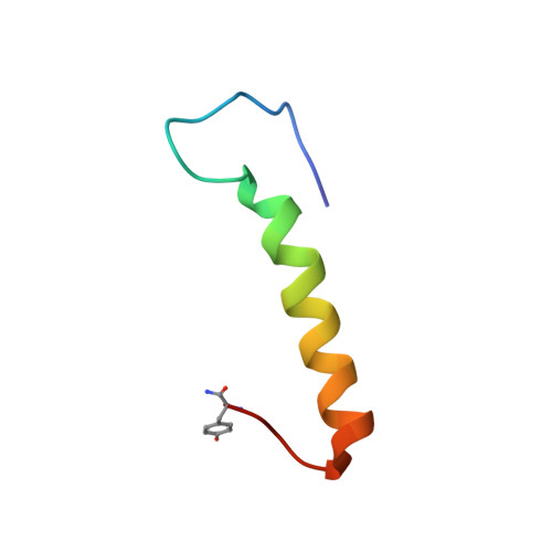
Bovine pancreatic polypeptide (bPP) undergoes significant changes in conformation and dynamics upon binding to DPC micelles.
Lerch, M., Gafner, V., Bader, R., Christen, B., Folkers, G., Zerbe, O.(2002) J Mol Biology 322: 1117-1133
- PubMed: 12367532 Search on PubMed
- DOI: https://doi.org/10.1016/s0022-2836(02)00889-6
- Primary Citation Related Structures:
1LJV - PubMed Abstract:
The pancreatic polypeptide (PP), a 36-residue, C-terminally amidated polypeptide hormone is a member of the neuropeptide Y (NPY) family. Here, we have studied the structure and dynamics of bovine pancreatic polypeptide (bPP) when bound to DPC-micelles as a membrane-mimicking model as well as the dynamics of bPP in solution. The comparison of structure and dynamics of bPP in both states reveals remarkable differences. The overall correlation time of 5.08ns derived from the 15N relaxation data proves unambiguously that bPP in solution exists as a dimer. Therein, intermolecular as well as intramolecular hydrophobic interactions from residues of both the amphiphilic helix and of the back-folded N terminus contribute to the stability of the PP fold. The overall rigidity is well-reflected in positive values for the heteronuclear NOE for residues 4-34. The membrane-bound species displays a partitioning into a more flexible N-terminal region and a well-defined alpha-helical region comprising residues 17-31. The average RMSD value for residues 17-31 is 0.22(+/-0.09)A. The flexibility of the N terminus is compatible with negative values of the heteronuclear NOE observed for the N-terminal residues 4-12 and low values of the generalized order parameter S(2). The membrane-peptide interface was investigated by micelle-integrating spin-labels and H,2H exchange measurements. It is formed by those residues which make contacts between the C-terminal alpha-helix and the polyproline helix. In contrast to pNPY, also residues from the N terminus display spatial proximity to the membrane interface. Furthermore, the orientation of the C terminus, that presumably contains residues involved in receptor binding, is different in the two environments. We speculate that this pre-positioning of residues could be an important requirement for receptor activation. Moreover, we doubt that the PP fold is of functional relevance for binding at the Y(4) receptor.
- Department of Applied BioSciences, Institute of Pharmaceutical Sciences, ETH Zurich, Winterthurerstrasse 190, CH 8057 Zurich, Switzerland.
Organizational Affiliation: