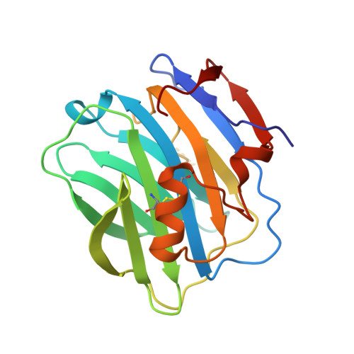
The three-dimensional structure of calcium-depleted human C-reactive protein from perfectly twinned crystals.
Ramadan, M.A., Shrive, A.K., Holden, D., Myles, D.A., Volanakis, J.E., DeLucas, L.J., Greenhough, T.J.(2002) Acta Crystallogr D Biol Crystallogr 58: 992-1001
- PubMed: 12037301
- DOI: https://doi.org/10.1107/s0907444902005693
- Primary Citation of Related Structures:
1LJ7 - PubMed Abstract:
C-reactive protein is a member of the pentraxin family of oligomeric serum proteins which has been conserved through evolution, homologues having been found in every species in which they have been sought. Human C-reactive protein (hCRP) is the classical acute-phase reactant produced in large amounts in response to tissue damage and inflammation and is used almost universally as a clinical indicator of infection and inflammation. The role of hCRP in host defence and the calcium-dependent ligand-binding specificity of hCRP for phosphocholine moieties have long been recognized. In order to clarify the structural rearrangements associated with calcium binding, the reported affinity of calcium-depleted hCRP for polycations and other ligands, and the role of calcium in protection against denaturation and proteolysis, the structure of calcium-depleted hCRP has been determined by X-ray crystallography. Crystals of calcium-depleted hCRP are invariably twinned and those suitable for analysis are merohedral type II twins of point group 4 single crystals. The structure has been solved by molecular replacement using the calcium-bound hCRP structure [Shrive et al. (1996), Nature Struct. Biol. 3, 346-354]. It reveals two independent pentamers which form a face-to-face decamer across a dyad near-parallel to the twinning twofold axis. Cycles of intensity deconvolution, density modification (tenfold NCS) and model building, eventually including refinement, give a final R factor of 0.19 (R(free) = 0.20). Despite poor definition in some areas arising from the limited resolution of the data and from the twinning and disorder, the structure reveals the probable mode of twinning and the conformational changes, localized in one of the calcium-binding loops, which accompany calcium binding.
- School of Life Sciences, Keele University, Staffordshire ST5 5BG, England.
Organizational Affiliation:


