1LEM | pdb_00001lem

THE MONOSACCHARIDE BINDING SITE OF LENTIL LECTIN: AN X-RAY AND MOLECULAR MODELLING STUDY


Experimental Data Snapshot

  • Method: X-RAY DIFFRACTION
  • Resolution: 3.00 Å
  • R-Value Work: 
    0.206 (Depositor) 
  • R-Value Observed: 
    0.206 (Depositor) 

wwPDB Validation 3D Report Full Report

Validation slider image for 1LEM

This is version 1.5 of the entry. See complete history

Literature

The monosaccharide binding site of lentil lectin: an X-ray and molecular modelling study.

Loris, R.Casset, F.Bouckaert, J.Pletinckx, J.Dao-Thi, M.H.Poortmans, F.Imberty, A.Perez, S.Wyns, L.

(1994) Glycoconj J 11: 507-517

  • DOI: https://doi.org/10.1007/BF00731301
  • Primary Citation Related Structures: 
    1LEM

  • PubMed Abstract: 

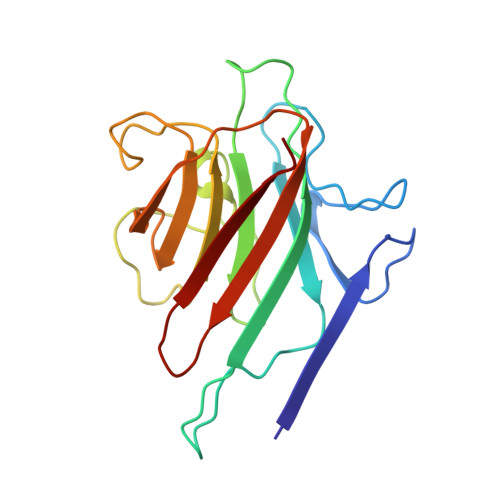
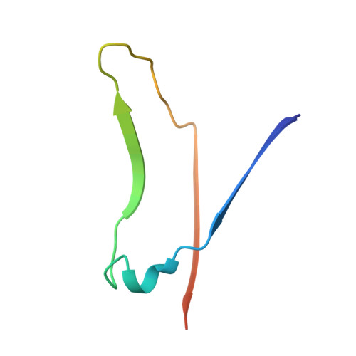
    The X-ray crystal structure of lentil lectin in complex with alpha-D-glucopyranose has been determined by molecular replacement and refined to an R-value of 0.20 at 3.0 A resolution. The glucose interacts with the protein in a manner similar to that found in the mannose complexes of concanavalin A, pea lectin and isolectin I from Lathyrus ochrus. The complex is stabilized by a network of hydrogen bonds involving the carbohydrate oxygens O6, O4, O3 and O5. In addition, the alpha-D-glucopyranose residue makes van der Waals contacts with the protein, involving the phenyl ring of Phe123 beta. The overall structure of lentil lectin, at this resolution, does not differ significantly from the highly refined structures of the uncomplexed lectin. Molecular docking studies were performed with mannose and its 2-O and 3-O-m-nitro-benzyl derivatives to explain their high affinity binding. The interactions of the modelled mannose with lentil lectin agree well with those observed experimentally for the protein-carbohydrate complex. The highly flexible Me-2-O-(m-nitro-benzyl)-alpha-D-mannopyranoside and Me-3-O-(m-nitro-benzyl)-alpha-D-mannopyranoside become conformationally restricted upon binding to lentil lectin. For best orientations of the two substrates in the combining site, the loss of entropy is accompanied by the formation of a strong hydrogen bond between the nitro group and one amino acid, Gly97 beta and Asn125 beta, respectively, along with the establishment of van der Waals interactions between the benzyl group and the aromatic amino acids Tyr100 beta and Trp128 beta.


  • Organizational Affiliation
    • Laboratorium voor Ultrastructuur, Vrije Universiteit Brussel, Sint-Genesius-Rode, Belgium.

Macromolecule Content 

  • Total Structure Weight: 25.9 kDa 
  • Atom Count: 1,833 
  • Modeled Residue Count: 228 
  • Deposited Residue Count: 233 
  • Unique protein chains: 2

Macromolecules

Find similar proteins by:|  3D Structure
Entity ID: 1
MoleculeChains  Sequence LengthOrganismDetailsImage
LECTIN181Lens culinarisMutation(s): 0 
UniProt
Find proteins for P02870 (Lens culinaris)
Explore P02870 
Go to UniProtKB:  P02870
Entity Groups
Sequence Clusters30% Identity50% Identity70% Identity90% Identity95% Identity100% Identity
UniProt GroupP02870
Sequence Annotations
Expand
Reference Sequence
Find similar proteins by:|  3D Structure
Entity ID: 2
MoleculeChains  Sequence LengthOrganismDetailsImage
LECTIN52Lens culinarisMutation(s): 0 
UniProt
Find proteins for P02870 (Lens culinaris)
Explore P02870 
Go to UniProtKB:  P02870
Entity Groups
Sequence Clusters30% Identity50% Identity70% Identity90% Identity95% Identity100% Identity
UniProt GroupP02870
Sequence Annotations
Expand
Reference Sequence

Small Molecules

Ligands 3 Unique
IDChains Name / Formula / InChI Key2D Diagram3D Interactions
GLC

Query on GLC


Download:Ideal Coordinates CCD File
C [auth A]alpha-D-glucopyranose
C6 H12 O6
WQZGKKKJIJFFOK-DVKNGEFBSA-N
MN

Query on MN


Download:Ideal Coordinates CCD File
E [auth A]MANGANESE (II) ION
Mn
WAEMQWOKJMHJLA-UHFFFAOYSA-N
CA

Query on CA


Download:Ideal Coordinates CCD File
D [auth A]CALCIUM ION
Ca
BHPQYMZQTOCNFJ-UHFFFAOYSA-N

Experimental Data & Validation

Experimental Data

  • Method: X-RAY DIFFRACTION
  • Resolution: 3.00 Å
  • R-Value Work:  0.206 (Depositor) 
  • R-Value Observed: 0.206 (Depositor) 
Space Group: P 65 2 2
Unit Cell:
Length ( Å )Angle ( ˚ )
a = 85.71α = 90
b = 85.71β = 90
c = 165.38γ = 120
Software Package:
Software NamePurpose
X-PLORmodel building
X-PLORrefinement
X-PLORphasing

Structure Validation

View Full Validation Report


Entry History 

Deposition Data

Revision History  (Full details and data files)

  • Version 1.0: 1994-01-31
    Type: Initial release
  • Version 1.1: 2008-03-03
    Changes: Version format compliance
  • Version 1.2: 2011-07-13
    Changes: Version format compliance
  • Version 1.3: 2017-11-29
    Changes: Derived calculations, Other
  • Version 1.4: 2020-07-29
    Type: Remediation
    Reason: Carbohydrate remediation
    Changes: Data collection, Derived calculations, Structure summary
  • Version 1.5: 2024-02-14
    Changes: Data collection, Database references, Structure summary