1LE8 | pdb_00001le8

Crystal Structure of the MATa1/MATalpha2-3A Heterodimer Bound to DNA Complex


Experimental Data Snapshot

  • Method: X-RAY DIFFRACTION
  • Resolution: 2.30 Å
  • R-Value Free: 
    0.299 (Depositor), 0.296 (DCC) 
  • R-Value Work: 
    0.256 (Depositor), 0.254 (DCC) 

wwPDB Validation 3D Report Full Report

Validation slider image for 1LE8

This is version 1.4 of the entry. See complete history

Literature

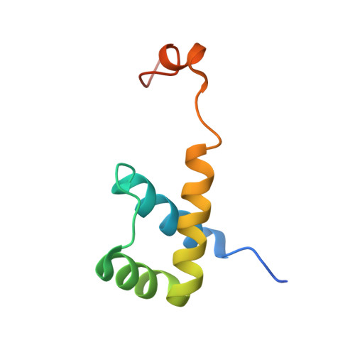
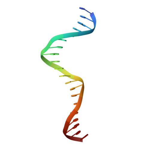
Structural and Thermodynamic Characterization of the DNA Binding Properties of a Triple Alanine Mutant of MATalpha2

Ke, A.Mathias, J.R.Vershon, A.K.Wolberger, C.

(2002) Structure 10: 961-971

  • DOI: https://doi.org/10.1016/s0969-2126(02)00790-6
  • Primary Citation Related Structures: 
    1LE8

  • PubMed Abstract: 

    Triply mutated MATalpha2 protein, alpha2-3A, in which all three major groove-contacting residues are mutated to alanine, is defective in binding DNA alone or in complex with Mcm1 yet binds with MATa1 with near wild-type affinity and specificity. To gain insight into this unexpected behavior, we determined the crystal structure of the a1/alpha2-3A/DNA complex. The structure shows that the triple mutation causes a collapse of the alpha2-3A/DNA interface that results in a reorganized set of alpha2-3A/DNA contacts, thereby enabling the mutant protein to recognize the wild-type DNA sequence. Isothermal titration calorimetry measurements reveal that a much more favorable entropic component stabilizes the a1/alpha2-3A/DNA complex than the alpha2-3A/DNA complex. The combined structural and thermodynamic studies provide an explanation of how partner proteins influence the sequence specificity of a DNA binding protein.


  • Organizational Affiliation
    • Department of Biophysics and Biophysical Chemistry, John Hopkins University, 725 North Wolfe Street, Baltimore, MD 21205, USA.

Macromolecule Content 

  • Total Structure Weight: 28.17 kDa 
  • Atom Count: 1,909 
  • Modeled Residue Count: 167 
  • Deposited Residue Count: 176 
  • Unique protein chains: 2
  • Unique nucleic acid chains: 2

Macromolecules


Find similar proteins by:|  3D Structure
Entity ID: 3
MoleculeChains  Sequence LengthOrganismDetailsImage
MATING-TYPE PROTEIN A-1C [auth A]53N/AMutation(s): 0 
UniProt
Find proteins for P0CY10 (Saccharomyces cerevisiae)
Explore P0CY10 
Go to UniProtKB:  P0CY10
Entity Groups
Sequence Clusters30% Identity50% Identity70% Identity90% Identity95% Identity100% Identity
UniProt GroupP0CY10
Sequence Annotations
Expand
Reference Sequence
Find similar proteins by:|  3D Structure
Entity ID: 4
MoleculeChains  Sequence LengthOrganismDetailsImage
Mating-type protein alpha-2D [auth B]83Saccharomyces cerevisiaeMutation(s): 3 
Gene Names: MATalpha2
UniProt
Find proteins for P0CY08 (Saccharomyces cerevisiae (strain ATCC 204508 / S288c))
Explore P0CY08 
Go to UniProtKB:  P0CY08
Entity Groups
Sequence Clusters30% Identity50% Identity70% Identity90% Identity95% Identity100% Identity
UniProt GroupP0CY08
Sequence Annotations
Expand
Reference Sequence
Find similar nucleic acids by:  Sequence
Entity ID: 1
MoleculeChains LengthOrganismImage
5'-D(*AP*CP*AP*TP*GP*TP*AP*AP*AP*AP*AP*TP*TP*TP*AP*CP*AP*TP*CP*A)-3'A [auth C]20N/A
Sequence Annotations
Expand
Reference Sequence
Find similar nucleic acids by:  Sequence
Entity ID: 2
MoleculeChains LengthOrganismImage
5'-D(*TP*TP*GP*AP*TP*GP*TP*AP*AP*AP*TP*TP*TP*TP*TP*AP*CP*AP*TP*G)-3'B [auth D]20N/A
Sequence Annotations
Expand
Reference Sequence

Experimental Data & Validation

Experimental Data

  • Method: X-RAY DIFFRACTION
  • Resolution: 2.30 Å
  • R-Value Free:  0.299 (Depositor), 0.296 (DCC) 
  • R-Value Work:  0.256 (Depositor), 0.254 (DCC) 
Space Group: P 65
Unit Cell:
Length ( Å )Angle ( ˚ )
a = 54.17α = 90
b = 54.17β = 90
c = 164.23γ = 120
Software Package:
Software NamePurpose
MOSFLMdata reduction
SCALEPACKdata scaling
AMoREphasing
CNSrefinement

Structure Validation

View Full Validation Report


Entry History 

Deposition Data

Revision History  (Full details and data files)

  • Version 1.0: 2002-05-03
    Type: Initial release
  • Version 1.1: 2008-04-28
    Changes: Version format compliance
  • Version 1.2: 2011-07-13
    Changes: Version format compliance
  • Version 1.3: 2021-10-27
    Changes: Database references
  • Version 1.4: 2024-02-14
    Changes: Data collection