Structure function relationship of reduced cytochrome c probed by complete solution structure determination in 30% acetonitrile/water solution
Sivakolundu, S.G., Mabrouk, P.A.(2003) J Biol Inorg Chem 8: 527-539
- PubMed: 12764601 Search on PubMed
- DOI: https://doi.org/10.1007/s00775-002-0437-0
- Primary Citation Related Structures:
1LC1, 1LC2 - PubMed Abstract:
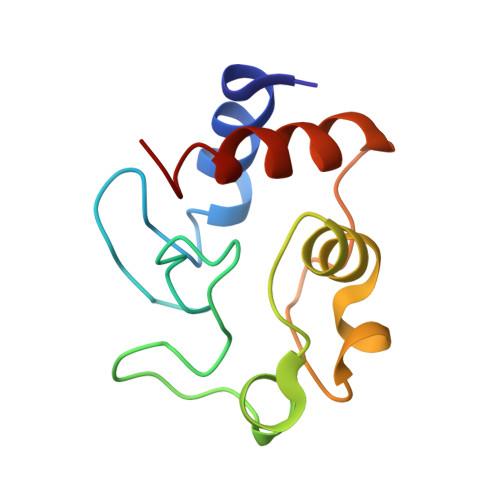
The complete solution structure of ferrocytochrome c in 30% acetonitrile/70% water has been determined using high-field 1D and 2D (1)H NMR methods and deposited in the Protein Data Bank with codes 1LC1 and 1LC2. This is the first time a complete solution protein structure has been determined for a protein in nonaqueous media. Ferrocyt c retains a native protein secondary structure (five alpha-helices and two omega loops) in 30% acetonitrile. H18 and M80 residues are the axial heme ligands, as in aqueous solution. Residues believed to be axial heme ligands in the alkaline-like conformers of ferricyt c, specifically H33 and K72, are positioned close to the heme iron. The orientations of both heme propionates are markedly different in 30% acetonitrile/70% water. Comparative structural analysis of reduced cyt c in 30% acetonitrile/70% water solution with cyt c in different environments has given new insight into the cyt c folding mechanism, the electron transfer pathway, and cell apoptosis.
- Department of Chemistry and Chemical Biology, Northeastern University, Boston, MA 02115, USA.
Organizational Affiliation: