1L2U | pdb_00001l2u

Orotidine 5'-monophosphate decarboxylase from E. coli


Experimental Data Snapshot

  • Method: X-RAY DIFFRACTION
  • Resolution: 2.50 Å
  • R-Value Free: 
    0.211 (Depositor), 0.185 (DCC) 
  • R-Value Work: 
    0.159 (Depositor), 0.143 (DCC) 
  • R-Value Observed: 
    0.162 (Depositor) 

Starting Model: experimental
View more details

wwPDB Validation 3D Report Full Report

Validation slider image for 1L2U

This is version 1.4 of the entry. See complete history

Literature

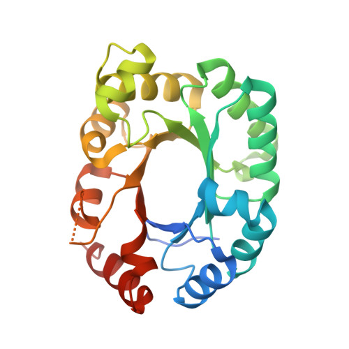
Substrate binding induces domain movements in orotidine 5'-monophosphate decarboxylase

Harris, P.Poulsen, J.C.Jensen, K.F.Larsen, S.

(2002) J Mol Biology 18: 1019-1029

  • DOI: https://doi.org/10.1016/S0022-2836(02)00200-0
  • Primary Citation Related Structures: 
    1L2U

  • PubMed Abstract: 

    Orotidine 5'-monophosphate decarboxylase (ODCase) catalyses the decarboxylation of orotidine 5'-monophosphate to uridine 5'-monophosphate (UMP). We have earlier determined the structure of ODCase from Escherichia coli complexed with the inhibitor 1-(5'-phospho-beta-d-ribofuranosyl)barbituric acid (BMP); here we present the 2.5 A structure of the uncomplexed apo enzyme, determined from twinned crystals. A structural analysis and comparison of the two structures of the E. coli enzyme show that binding of the inhibitor is accompanied by significant domain movements of approximately 12 degrees around a hinge that crosses the active site. Hence, the ODCase dimer, which contains two active sites, may be divided in three domains: a central domain that is fixed, and two lids which independently move 12 degrees upon binding. Corresponding analyses, presented herein, of the two Saccharomyces cerevisiae ODCase structures (with and without BMP) and the Methanobacterium thermoautotrophicum ODCase structures (with and without 6-aza UMP) show very similar, but somewhat smaller domain movements. The domain movements seem to be initiated by the phosphoryl binding to the enzyme and can explain why the binding of the phosphoryl group is essential for the catalytic function.


  • Organizational Affiliation
    • Centre for Crystallographic Studies, Department of Chemistry, University of Copenhagen, Universitetsparken 5, Denmark.

Macromolecule Content 

  • Total Structure Weight: 52.76 kDa 
  • Atom Count: 3,512 
  • Modeled Residue Count: 447 
  • Deposited Residue Count: 490 
  • Unique protein chains: 1

Macromolecules

Find similar proteins by:|  3D Structure
Entity ID: 1
MoleculeChains  Sequence LengthOrganismDetailsImage
Orotidine 5'-phosphate decarboxylase
A, B
245Escherichia coliMutation(s): 0 
EC: 4.1.1.23
UniProt
Find proteins for P08244 (Escherichia coli (strain K12))
Explore P08244 
Go to UniProtKB:  P08244
Entity Groups
Sequence Clusters30% Identity50% Identity70% Identity90% Identity95% Identity100% Identity
UniProt GroupP08244
Sequence Annotations
Expand
Reference Sequence

Experimental Data & Validation

Experimental Data

  • Method: X-RAY DIFFRACTION
  • Resolution: 2.50 Å
  • R-Value Free:  0.211 (Depositor), 0.185 (DCC) 
  • R-Value Work:  0.159 (Depositor), 0.143 (DCC) 
  • R-Value Observed: 0.162 (Depositor) 
Space Group: P 32 2 1
Unit Cell:
Length ( Å )Angle ( ˚ )
a = 142.69α = 90
b = 142.69β = 90
c = 105.37γ = 120
Software Package:
Software NamePurpose
DENZOdata reduction
SCALEPACKdata scaling
AMoREphasing
SHELXL-97refinement

Structure Validation

View Full Validation Report


Entry History 

Deposition Data

Revision History  (Full details and data files)

  • Version 1.0: 2002-03-13
    Type: Initial release
  • Version 1.1: 2008-04-28
    Changes: Version format compliance
  • Version 1.2: 2011-07-13
    Changes: Version format compliance
  • Version 1.3: 2018-03-07
    Changes: Data collection
  • Version 1.4: 2023-08-16
    Changes: Data collection, Database references, Refinement description