1KUJ

Crystal structure of Jacalin complexed with 1-O-methyl-alpha-D-mannose


Experimental Data Snapshot

  • Method: X-RAY DIFFRACTION
  • Resolution: 2.00 Å
  • R-Value Free: 0.235 
  • R-Value Work: 0.191 
  • R-Value Observed: 0.191 

Starting Model: experimental
View more details

wwPDB Validation   3D Report Full Report


Ligand Structure Quality Assessment 


This is version 1.4 of the entry. See complete history


Literature

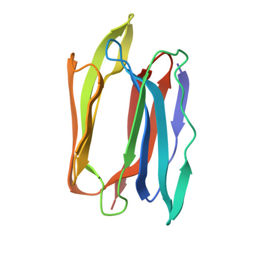
Structural basis for the unusual carbohydrate-binding specificity of jacalin towards galactose and mannose.

Bourne, Y.Astoul, C.H.Zamboni, V.Peumans, W.J.Menu-Bouaouiche, L.Van Damme, E.J.Barre, A.Rouge, P.

(2002) Biochem J 364: 173-180

  • DOI: https://doi.org/10.1042/bj3640173
  • Primary Citation of Related Structures:  
    1KU8, 1KUJ

  • PubMed Abstract: 

    Evidence is presented that the specificity of jacalin, the seed lectin from jack fruit (Artocarpus integrifolia), is not directed exclusively against the T-antigen disaccharide Galbeta1,3GalNAc, lactose and galactose, but also against mannose and oligomannosides. Biochemical analyses based on surface-plasmon-resonance measurements, combined with the X-ray-crystallographic determination of the structure of a jacalin-alpha-methyl-mannose complex at 2 A resolution, demonstrated clearly that jacalin is fully capable of binding mannose. Besides mannose, jacalin also interacts readily with glucose, N-acetylneuraminic acid and N-acetylmuramic acid. Structural analyses demonstrated that the relatively large size of the carbohydrate-binding site enables jacalin to accommodate monosaccharides with different hydroxyl conformations and provided unambiguous evidence that the beta-prism structure of jacalin is a sufficiently flexible structural scaffold to confer different carbohydrate-binding specificities to a single lectin.


  • Organizational Affiliation

    AFMB, UMR-CNRS 6098, 31 Chemin Joseph Aiguier, 13402 Marseille Cedex 20, France.


Macromolecules
Find similar proteins by:  (by identity cutoff)  |  3D Structure
Entity ID: 1
MoleculeChains Sequence LengthOrganismDetailsImage
JACALIN ALPHA CHAIN
A, C, E, G
133Artocarpus integerMutation(s): 0 
UniProt
Find proteins for P18670 (Artocarpus integer)
Explore P18670 
Go to UniProtKB:  P18670
Entity Groups  
Sequence Clusters30% Identity50% Identity70% Identity90% Identity95% Identity100% Identity
UniProt GroupP18670
Sequence Annotations
Expand
  • Reference Sequence

Find similar proteins by:  Sequence   |   3D Structure  

Entity ID: 2
MoleculeChains Sequence LengthOrganismDetailsImage
JACALIN BETA CHAIN
B, D, F, H
18Artocarpus integerMutation(s): 0 
UniProt
Find proteins for P18671 (Artocarpus integer)
Explore P18671 
Go to UniProtKB:  P18671
Entity Groups  
Sequence Clusters30% Identity50% Identity70% Identity90% Identity95% Identity100% Identity
UniProt GroupP18671
Sequence Annotations
Expand
  • Reference Sequence
Experimental Data & Validation

Experimental Data

  • Method: X-RAY DIFFRACTION
  • Resolution: 2.00 Å
  • R-Value Free: 0.235 
  • R-Value Work: 0.191 
  • R-Value Observed: 0.191 
  • Space Group: P 1 21 1
Unit Cell:
Length ( Å )Angle ( ˚ )
a = 58.795α = 90
b = 82.612β = 106.8
c = 63.482γ = 90
Software Package:
Software NamePurpose
CNSrefinement
DENZOdata reduction
CCP4data scaling
CNSphasing

Structure Validation

View Full Validation Report


Ligand Structure Quality Assessment 


Entry History 

Deposition Data

Revision History  (Full details and data files)

  • Version 1.0: 2002-06-19
    Type: Initial release
  • Version 1.1: 2008-04-27
    Changes: Version format compliance
  • Version 1.2: 2011-07-13
    Changes: Version format compliance
  • Version 1.3: 2020-07-29
    Type: Remediation
    Reason: Carbohydrate remediation
    Changes: Data collection, Derived calculations, Structure summary
  • Version 1.4: 2023-08-16
    Changes: Data collection, Database references, Refinement description, Structure summary