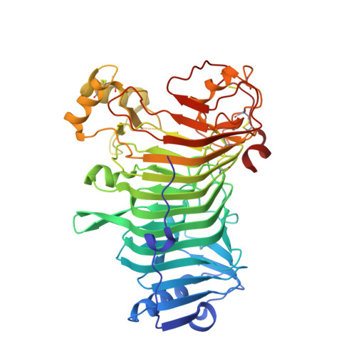
1KTW

IOTA-CARRAGEENASE COMPLEXED TO IOTA-CARRAGEENAN FRAGMENTS


Experimental Data Snapshot

  • Method: X-RAY DIFFRACTION
  • Resolution: 2.00 Å
  • R-Value Free: 0.238 
  • R-Value Work: 0.203 

wwPDB Validation   3D Report Full Report


This is version 2.1 of the entry. See complete history


Literature

The Structural Bases of the Processive Degradation of iota-Carrageenan, a Main Cell Wall Polysaccharide of Red Algae.

Michel, G.Helbert, W.Kahn, R.Dideberg, O.Kloareg, B.

(2003) J Mol Biol 334: 421-433

  • DOI: https://doi.org/10.1016/j.jmb.2003.09.056
  • Primary Citation of Related Structures:  
    1KTW

  • PubMed Abstract: 

    iota-Carrageenans are sulfated 1,3-alpha-1,4-beta-galactans from the cell walls of red algae, which auto-associate into crystalline fibers made of aggregates of double-stranded helices. iota-Carrageenases, which constitute family 82 of glycoside hydrolases, fold into a right-handed beta-helix. Here, the structure of Alteromonas fortis iota-carrageenase bound to iota-carrageenan fragments was solved at 2.0A resolution (PDB 1KTW). The enzyme holds a iota-carrageenan tetrasaccharide (subsites +1 to +4) and a disaccharide (subsites -3, -4), thus providing the first direct determination of a 3D structure of iota-carrageenan. Electrostatic interactions between basic protein residues and the sulfate substituents of the polysaccharide chain dominate iota-carrageenan recognition. Glu245 and Asp247 are the proton donor and the base catalyst, respectively. C-terminal domain A, which was highly flexible in the native enzyme structure, adopts a alpha/beta-fold, also found in DNA/RNA-binding domains. In the substrate-enzyme complex, this polyanion-binding module shifts toward the beta-helix groove, forming a tunnel. Thus, from an open conformation which allows for the initial endo-attack of iota-carrageenan chains, the enzyme switches to a closed-tunnel form, consistent with its highly processive character, as seen from the electron-microscopy analysis of the degradation of iota-carrageenan fibers.


  • Organizational Affiliation

    Végétaux Marins et Biomolécules, UMR 7139 (CNRS/UPMC/Laboratories Goëmar), Station Biologique de Roscoff, Place Georges Teissier, BP 74, 29682 Roscoff Cedex, Brittany, France.kloareg@sb-rosecoff.ff


Macromolecules
Find similar proteins by:  (by identity cutoff)  |  3D Structure
Entity ID: 1
MoleculeChains Sequence LengthOrganismDetailsImage
IOTA-CARRAGEENASE
A, B
464Alteromonas macleodiiMutation(s): 0 
EC: 3.2.1.157
UniProt
Find proteins for Q9F5I8 (Alteromonas macleodii)
Explore Q9F5I8 
Go to UniProtKB:  Q9F5I8
Entity Groups  
Sequence Clusters30% Identity50% Identity70% Identity90% Identity95% Identity100% Identity
UniProt GroupQ9F5I8
Sequence Annotations
Expand
  • Reference Sequence
Oligosaccharides

Help

Entity ID: 2
MoleculeChains Length2D Diagram Glycosylation3D Interactions
3,6-anhydro-2-O-sulfo-alpha-D-galactopyranose-(1-3)-4-O-sulfo-beta-D-galactopyranose
C
2N/A
Glycosylation Resources
GlyTouCan:  G58187OK
GlyCosmos:  G58187OK
Entity ID: 3
MoleculeChains Length2D Diagram Glycosylation3D Interactions
3,6-anhydro-2-O-sulfo-alpha-D-galactopyranose-(1-3)-4-O-sulfo-beta-D-galactopyranose-(1-4)-3,6-anhydro-2-O-sulfo-alpha-D-galactopyranose-(1-3)-4-O-sulfo-beta-D-galactopyranose
D
4N/A
Glycosylation Resources
GlyTouCan:  G24165XP
GlyCosmos:  G24165XP
Small Molecules
Ligands 3 Unique
IDChains Name / Formula / InChI Key2D Diagram3D Interactions
CA
Query on CA

Download Ideal Coordinates CCD File 
E [auth A]
F [auth A]
G [auth A]
H [auth A]
I [auth A]
E [auth A],
F [auth A],
G [auth A],
H [auth A],
I [auth A],
M [auth B],
N [auth B],
O [auth B],
P [auth B],
Q [auth B]
CALCIUM ION
Ca
BHPQYMZQTOCNFJ-UHFFFAOYSA-N
CL
Query on CL

Download Ideal Coordinates CCD File 
L [auth A],
T [auth B]
CHLORIDE ION
Cl
VEXZGXHMUGYJMC-UHFFFAOYSA-M
NA
Query on NA

Download Ideal Coordinates CCD File 
J [auth A],
K [auth A],
R [auth B],
S [auth B]
SODIUM ION
Na
FKNQFGJONOIPTF-UHFFFAOYSA-N
Experimental Data & Validation

Experimental Data

  • Method: X-RAY DIFFRACTION
  • Resolution: 2.00 Å
  • R-Value Free: 0.238 
  • R-Value Work: 0.203 
  • Space Group: P 1 21 1
Unit Cell:
Length ( Å )Angle ( ˚ )
a = 54.73α = 90
b = 122.31β = 90.99
c = 91.941γ = 90
Software Package:
Software NamePurpose
DENZOdata reduction
SCALEPACKdata scaling
AMoREphasing
CNSrefinement

Structure Validation

View Full Validation Report


Entry History 

Deposition Data

Revision History  (Full details and data files)

  • Version 1.0: 2003-06-10
    Type: Initial release
  • Version 1.1: 2008-04-27
    Changes: Version format compliance
  • Version 1.2: 2011-07-13
    Changes: Version format compliance
  • Version 2.0: 2020-07-29
    Type: Remediation
    Reason: Carbohydrate remediation
    Changes: Advisory, Atomic model, Data collection, Derived calculations, Structure summary
  • Version 2.1: 2023-08-16
    Changes: Advisory, Data collection, Database references, Refinement description, Structure summary