1KTQ | pdb_00001ktq

DNA POLYMERASE


Experimental Data Snapshot

  • Method: X-RAY DIFFRACTION
  • Resolution: 2.50 Å
  • R-Value Free: 
    0.262 (Depositor) 
  • R-Value Work: 
    0.198 (Depositor) 
  • R-Value Observed: 
    0.198 (Depositor) 

wwPDB Validation 3D Report Full Report

Validation slider image for 1KTQ

This is version 1.4 of the entry. See complete history

Literature

Crystal structure of the large fragment of Thermus aquaticus DNA polymerase I at 2.5-A resolution: structural basis for thermostability.

Korolev, S.Nayal, M.Barnes, W.M.Di Cera, E.Waksman, G.

(1995) Proc Natl Acad Sci U S A 92: 9264-9268

  • DOI: https://doi.org/10.1073/pnas.92.20.9264
  • Primary Citation Related Structures: 
    1KTQ

  • PubMed Abstract: 

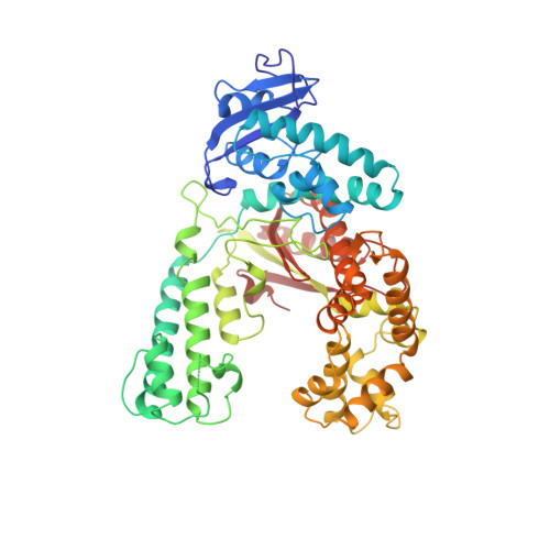
    The crystal structure of the large fragment of the Thermus aquaticus DNA polymerase (Klentaq1), determined at 2.5-A resolution, demonstrates a compact two-domain architecture. The C-terminal domain is identical in fold to the equivalent region of the Klenow fragment of Escherichia coli DNA polymerase I (Klenow pol I). Although the N-terminal domain of Klentaq1 differs greatly in sequence from its counterpart in Klenow pol I, it has clearly evolved from a common ancestor. The structure of Klentaq1 reveals the strategy utilized by this protein to maintain activity at high temperatures and provides the structural basis for future improvements of the enzyme.


  • Organizational Affiliation
    • Department of Biochemistry and Molecular Biophysics, Washington University School of Medicine, St. Louis, MO 63110, USA.

Macromolecule Content 

  • Total Structure Weight: 61.25 kDa 
  • Atom Count: 4,679 
  • Modeled Residue Count: 534 
  • Deposited Residue Count: 543 
  • Unique protein chains: 1

Macromolecules

Find similar proteins by:|  3D Structure
Entity ID: 1
MoleculeChains  Sequence LengthOrganismDetailsImage
DNA POLYMERASE I543Thermus aquaticusMutation(s): 0 
EC: 2.7.7.7
UniProt
Find proteins for P19821 (Thermus aquaticus)
Explore P19821 
Go to UniProtKB:  P19821
Entity Groups
Sequence Clusters30% Identity50% Identity70% Identity90% Identity95% Identity100% Identity
UniProt GroupP19821
Sequence Annotations
Expand
Reference Sequence

Experimental Data & Validation

Experimental Data

  • Method: X-RAY DIFFRACTION
  • Resolution: 2.50 Å
  • R-Value Free:  0.262 (Depositor) 
  • R-Value Work:  0.198 (Depositor) 
  • R-Value Observed: 0.198 (Depositor) 
Space Group: P 21 21 2
Unit Cell:
Length ( Å )Angle ( ˚ )
a = 109.4α = 90
b = 136.8β = 90
c = 45.6γ = 90
Software Package:
Software NamePurpose
X-PLORmodel building
X-PLORrefinement
R-AXISdata reduction
X-PLORphasing

Structure Validation

View Full Validation Report


Entry History 

Deposition Data

Revision History  (Full details and data files)

  • Version 1.0: 1996-11-08
    Type: Initial release
  • Version 1.1: 2008-03-24
    Changes: Version format compliance
  • Version 1.2: 2011-07-13
    Changes: Version format compliance
  • Version 1.3: 2018-03-21
    Changes: Data collection, Other
  • Version 1.4: 2024-02-14
    Changes: Data collection, Database references