1KQF

FORMATE DEHYDROGENASE N FROM E. COLI


Experimental Data Snapshot

  • Method: X-RAY DIFFRACTION
  • Resolution: 1.60 Å
  • R-Value Free: 0.195 
  • R-Value Work: 0.177 
  • R-Value Observed: 0.177 

wwPDB Validation   3D Report Full Report


Ligand Structure Quality Assessment 


This is version 1.5 of the entry. See complete history


Literature

Molecular basis of proton motive force generation: structure of formate dehydrogenase-N.

Jormakka, M.Tornroth, S.Byrne, B.Iwata, S.

(2002) Science 295: 1863-1868

  • DOI: https://doi.org/10.1126/science.1068186
  • Primary Citation of Related Structures:  
    1KQF, 1KQG

  • PubMed Abstract: 

    The structure of the membrane protein formate dehydrogenase-N (Fdn-N), a major component of Escherichia coli nitrate respiration, has been determined at 1.6 angstroms. The structure demonstrates 11 redox centers, including molybdopterin-guanine dinucleotides, five [4Fe-4S] clusters, two heme b groups, and a menaquinone analog. These redox centers are aligned in a single chain, which extends almost 90 angstroms through the enzyme. The menaquinone reduction site associated with a possible proton pathway was also characterized. This structure provides critical insights into the proton motive force generation by redox loop, a common mechanism among a wide range of respiratory enzymes.


  • Organizational Affiliation

    Division of Biomedical Sciences, Imperial College, London SW7 2AZ, UK.


Macromolecules
Find similar proteins by:  (by identity cutoff)  |  3D Structure
Entity ID: 1
MoleculeChains Sequence LengthOrganismDetailsImage
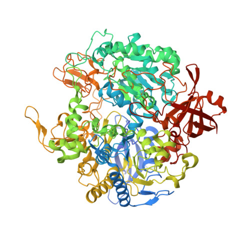
FORMATE DEHYDROGENASE, NITRATE-INDUCIBLE, MAJOR SUBUNIT1,015Escherichia coliMutation(s): 0 
EC: 1.2.1.2 (PDB Primary Data), 1.17.5.3 (UniProt)
Membrane Entity: Yes 
UniProt
Find proteins for P24183 (Escherichia coli (strain K12))
Explore P24183 
Go to UniProtKB:  P24183
Entity Groups  
Sequence Clusters30% Identity50% Identity70% Identity90% Identity95% Identity100% Identity
UniProt GroupP24183
Sequence Annotations
Expand
  • Reference Sequence
Find similar proteins by:  (by identity cutoff)  |  3D Structure
Entity ID: 2
MoleculeChains Sequence LengthOrganismDetailsImage
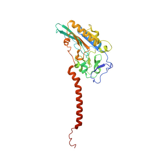
FORMATE DEHYDROGENASE, NITRATE-INDUCIBLE, IRON-SULFUR SUBUNIT294Escherichia coliMutation(s): 0 
EC: 1.2.1.2
Membrane Entity: Yes 
UniProt
Find proteins for P0AAJ3 (Escherichia coli (strain K12))
Explore P0AAJ3 
Go to UniProtKB:  P0AAJ3
Entity Groups  
Sequence Clusters30% Identity50% Identity70% Identity90% Identity95% Identity100% Identity
UniProt GroupP0AAJ3
Sequence Annotations
Expand
  • Reference Sequence
Find similar proteins by:  (by identity cutoff)  |  3D Structure
Entity ID: 3
MoleculeChains Sequence LengthOrganismDetailsImage
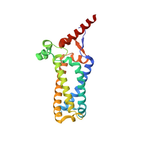
FORMATE DEHYDROGENASE, NITRATE-INDUCIBLE, CYTOCHROME B556(FDN) SUBUNIT217Escherichia coliMutation(s): 0 
EC: 1.2.1.2
Membrane Entity: Yes 
UniProt
Find proteins for P0AEK7 (Escherichia coli (strain K12))
Explore P0AEK7 
Go to UniProtKB:  P0AEK7
Entity Groups  
Sequence Clusters30% Identity50% Identity70% Identity90% Identity95% Identity100% Identity
UniProt GroupP0AEK7
Sequence Annotations
Expand
  • Reference Sequence
Small Molecules
Ligands 5 Unique
IDChains Name / Formula / InChI Key2D Diagram3D Interactions
CDL
Query on CDL

Download Ideal Coordinates CCD File 
N [auth C]CARDIOLIPIN
C81 H156 O17 P2
XVTUQDWPJJBEHJ-KZCWQMDCSA-L
MGD
Query on MGD

Download Ideal Coordinates CCD File 
F [auth A],
G [auth A]
2-AMINO-5,6-DIMERCAPTO-7-METHYL-3,7,8A,9-TETRAHYDRO-8-OXA-1,3,9,10-TETRAAZA-ANTHRACEN-4-ONE GUANOSINE DINUCLEOTIDE
C20 H26 N10 O13 P2 S2
VQAGYJCYOLHZDH-ILXWUORBSA-N
HEM
Query on HEM

Download Ideal Coordinates CCD File 
L [auth C],
M [auth C]
PROTOPORPHYRIN IX CONTAINING FE
C34 H32 Fe N4 O4
KABFMIBPWCXCRK-RGGAHWMASA-L
SF4
Query on SF4

Download Ideal Coordinates CCD File 
E [auth A],
H [auth B],
I [auth B],
J [auth B],
K [auth B]
IRON/SULFUR CLUSTER
Fe4 S4
LJBDFODJNLIPKO-UHFFFAOYSA-N
6MO
Query on 6MO

Download Ideal Coordinates CCD File 
D [auth A]MOLYBDENUM(VI) ION
Mo
HCNGUXXTNNIKCQ-UHFFFAOYSA-N
Experimental Data & Validation

Experimental Data

  • Method: X-RAY DIFFRACTION
  • Resolution: 1.60 Å
  • R-Value Free: 0.195 
  • R-Value Work: 0.177 
  • R-Value Observed: 0.177 
  • Space Group: P 21 3
Unit Cell:
Length ( Å )Angle ( ˚ )
a = 203α = 90
b = 203β = 90
c = 203γ = 90
Software Package:
Software NamePurpose
MLPHAREphasing
CNSrefinement
DENZOdata reduction
SCALEPACKdata scaling

Structure Validation

View Full Validation Report


Ligand Structure Quality Assessment 


Entry History 

Deposition Data

Revision History  (Full details and data files)

  • Version 1.0: 2002-03-15
    Type: Initial release
  • Version 1.1: 2008-04-27
    Changes: Version format compliance
  • Version 1.2: 2011-07-13
    Changes: Derived calculations, Version format compliance
  • Version 1.3: 2014-02-19
    Changes: Atomic model, Structure summary
  • Version 1.4: 2014-08-06
    Changes: Derived calculations, Structure summary
  • Version 1.5: 2024-02-14
    Changes: Data collection, Database references, Derived calculations