A mutant RNA pseudoknot that promotes ribosomal frameshifting in mouse mammary tumor virus.
Kang, H., Tinoco Jr., I.(1997) Nucleic Acids Res 25: 1943-1949
- PubMed: 9115361 Search on PubMedSearch on PubMed Central
- DOI: https://doi.org/10.1093/nar/25.10.1943
- Primary Citation Related Structures:
1KPD - PubMed Abstract:
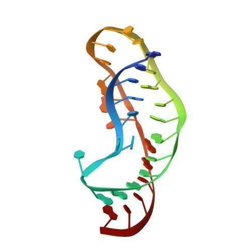
A single A-->G mutation that changes a potential A.U base pair to a G.U pair at the junction of the stems and loops of a non-frameshifting pseudoknot dramatically increases its frameshifting efficiency in mouse mammary tumor virus. The structure of the non-frameshifting pseudoknot APK has been found to be very different from that of pseudoknots that cause efficient frameshifting [Kang,H., Hines,J.V. and Tinoco,I. (1995) J. Mol. Biol. , 259, 135-147]. The 3-dimensional structure of the mutant pseudoknot was determined by restrained molecular dynamics based on NMR-derived interproton distance and torsion angle constraints. One striking feature of the mutant pseudoknot compared with the parent pseudoknot is that a G.U base pair forms at the top of stem 2, thus leaving only 1 nt at the junction of the two stems. The conformation is very different from that of the previously determined non-frameshifting parent pseudoknot, which lacks the A.U base pair at the top of the stem and has 2 nt between the stems. However, the conformation is quite similar to that of efficient frameshifting pseudoknots whose structures were previously determined by NMR. A single adenylate residue intervenes between the two stems and interrupts their coaxial stacking. This unpaired nucleotide produces a bent structure. The structural similarity among the efficient frameshifting pseudoknots indicates that a specific conformation is required for ribosomal frameshifting, further implying a specific interaction of the pseudoknot with the ribosome.
- Department of Chemistry, University of California, Berkeley CA 94720-1460, USA.
Organizational Affiliation: