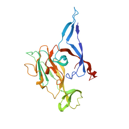
Crystal structure of a bacterial signal peptidase apoenzyme: implications for signal peptide binding and the Ser-Lys dyad mechanism
Paetzel, M., Dalbey, R.E., Strynadka, N.C.J.(2002) J Biological Chem 277: 9512-9519
- PubMed: 11741964
- DOI: https://doi.org/10.1074/jbc.M110983200
- Primary Citation of Related Structures:
1KN9 - PubMed Abstract:
We report here the x-ray crystal structure of a soluble catalytically active fragment of the Escherichia coli type I signal peptidase (SPase-(Delta2-75)) in the absence of inhibitor or substrate (apoenzyme). The structure was solved by molecular replacement and refined to 2.4 A resolution in a different space group (P4(1)2(1)2) from that of the previously published acyl-enzyme inhibitor-bound structure (P2(1)2(1)2) (Paetzel, M., Dalbey, R.E., and Strynadka, N.C.J. (1998) Nature 396, 186-190). A comparison with the acyl-enzyme structure shows significant side-chain and main-chain differences in the binding site and active site regions, which result in a smaller S1 binding pocket in the apoenzyme. The apoenzyme structure is consistent with SPase utilizing an unusual oxyanion hole containing one side-chain hydroxyl hydrogen (Ser-88 OgammaH) and one main-chain amide hydrogen (Ser-90 NH). Analysis of the apoenzyme active site reveals a potential deacylating water that was displaced by the inhibitor. It has been proposed that SPase utilizes a Ser-Lys dyad mechanism in the cleavage reaction. A similar mechanism has been proposed for the LexA family of proteases. A structural comparison of SPase and members of the LexA family of proteases reveals a difference in the side-chain orientation for the general base lysine, both of which are stabilized by an adjacent hydroxyl group. To gain insight into how signal peptidase recognizes its substrates, we have modeled a signal peptide into the binding site of SPase. The model is built based on the recently solved crystal structure of the analogous enzyme LexA (Luo, Y., Pfuetzner, R. A., Mosimann, S., Paetzel, M., Frey, E. A., Cherney, M., Kim, B., Little, J. W., and Strynadka, N. C. J. (2001) Cell 106, 1-10) with its bound cleavage site region.
- Department of Biochemistry and Molecular Biology, University of British Columbia, Vancouver, British Columbia, V6T 1Z3 Canada.
Organizational Affiliation:


