1KKQ | pdb_00001kkq

Crystal structure of the human PPAR-alpha ligand-binding domain in complex with an antagonist GW6471 and a SMRT corepressor motif


Experimental Data Snapshot

  • Method: X-RAY DIFFRACTION
  • Resolution: 3.00 Å
  • R-Value Free: 
    0.290 (Depositor) 
  • R-Value Work: 
    0.258 (Depositor) 
  • R-Value Observed: 
    0.290 (Depositor) 

Starting Model: experimental
View more details

wwPDB Validation 3D Report Full Report

Validation slider image for 1KKQ

This is version 1.6 of the entry. See complete history. 

Literature

Structural basis for antagonist-mediated recruitment of nuclear co-repressors by PPARalpha.

Xu, H.E., Stanley, T.B., Montana, V.G., Lambert, M.H., Shearer, B.G., Cobb, J.E., McKee, D.D., Galardi, C.M., Plunket, K.D., Nolte, R.T., Parks, D.J., Moore, J.T., Kliewer, S.A., Willson, T.M., Stimmel, J.B.

(2002) Nature 415: 813-817

  • DOI: https://doi.org/10.1038/415813a
  • Primary Citation Related Structures: 
    1KKQ

  • PubMed Abstract: 

    Repression of gene transcription by nuclear receptors is mediated by interactions with co-repressor proteins such as SMRT and N-CoR, which in turn recruit histone deacetylases to the chromatin. Aberrant interactions between nuclear receptors and co-repressors contribute towards acute promyelocytic leukaemia and thyroid hormone resistance syndrome. The binding of co-repressors to nuclear receptors occurs in the unliganded state, and can be stabilized by antagonists. Here we report the crystal structure of a ternary complex containing the peroxisome proliferator-activated receptor-alpha ligand-binding domain bound to the antagonist GW6471 and a SMRT co-repressor motif. In this structure, the co-repressor motif adopts a three-turn alpha-helix that prevents the carboxy-terminal activation helix (AF-2) of the receptor from assuming the active conformation. Binding of the co-repressor motif is further reinforced by the antagonist, which blocks the AF-2 helix from adopting the active position. Biochemical analyses and structure-based mutagenesis indicate that this mode of co-repressor binding is highly conserved across nuclear receptors.


  • Organizational Affiliation: 
    • Nuclear Receptor Discovery Research, GlaxoSmithKline, Research Triangle Park, NC 27709, USA. ex11957@gsk.com

Macromolecule Content 

  • Total Structure Weight: 133.21 kDa 
  • Atom Count: 9,606 
  • Modeled Residue Count: 1,152 
  • Deposited Residue Count: 1,152 
  • Unique protein chains: 2

Macromolecules

Find similar proteins by:|  3D Structure
Entity ID: 1
MoleculeChains  Sequence LengthOrganismDetailsImage
PEROXISOME PROLIFERATOR ACTIVATED RECEPTOR
A, B, C, D
269Homo sapiensMutation(s): 0 
Gene Names: human
UniProt & NIH Common Fund Data Resources
Find proteins for Q07869 (Homo sapiens)
Explore Q07869 
Go to UniProtKB:  Q07869
PHAROS:  Q07869
GTEx:  ENSG00000186951 
Entity Groups
Sequence Clusters30% Identity50% Identity70% Identity90% Identity95% Identity100% Identity
UniProt GroupQ07869
Sequence Annotations
Expand
Reference Sequence
Find similar proteins by:  Sequence   |   3D Structure  
Entity ID: 2
MoleculeChains  Sequence LengthOrganismDetailsImage
NUCLEAR RECEPTOR CO-REPRESSOR 2
E, F, G, H
19N/AMutation(s): 0 
UniProt & NIH Common Fund Data Resources
Find proteins for Q9Y618 (Homo sapiens)
Explore Q9Y618 
Go to UniProtKB:  Q9Y618
PHAROS:  Q9Y618
GTEx:  ENSG00000196498 
Entity Groups
Sequence Clusters30% Identity50% Identity70% Identity90% Identity95% Identity100% Identity
UniProt GroupQ9Y618
Sequence Annotations
Expand
Reference Sequence

Small Molecules

Ligands 1 Unique
IDChains Name / Formula / InChI Key2D Diagram3D Interactions
471

Query on 471


Download:Ideal Coordinates CCD File
I [auth A],
J [auth B],
K [auth C],
L [auth D]
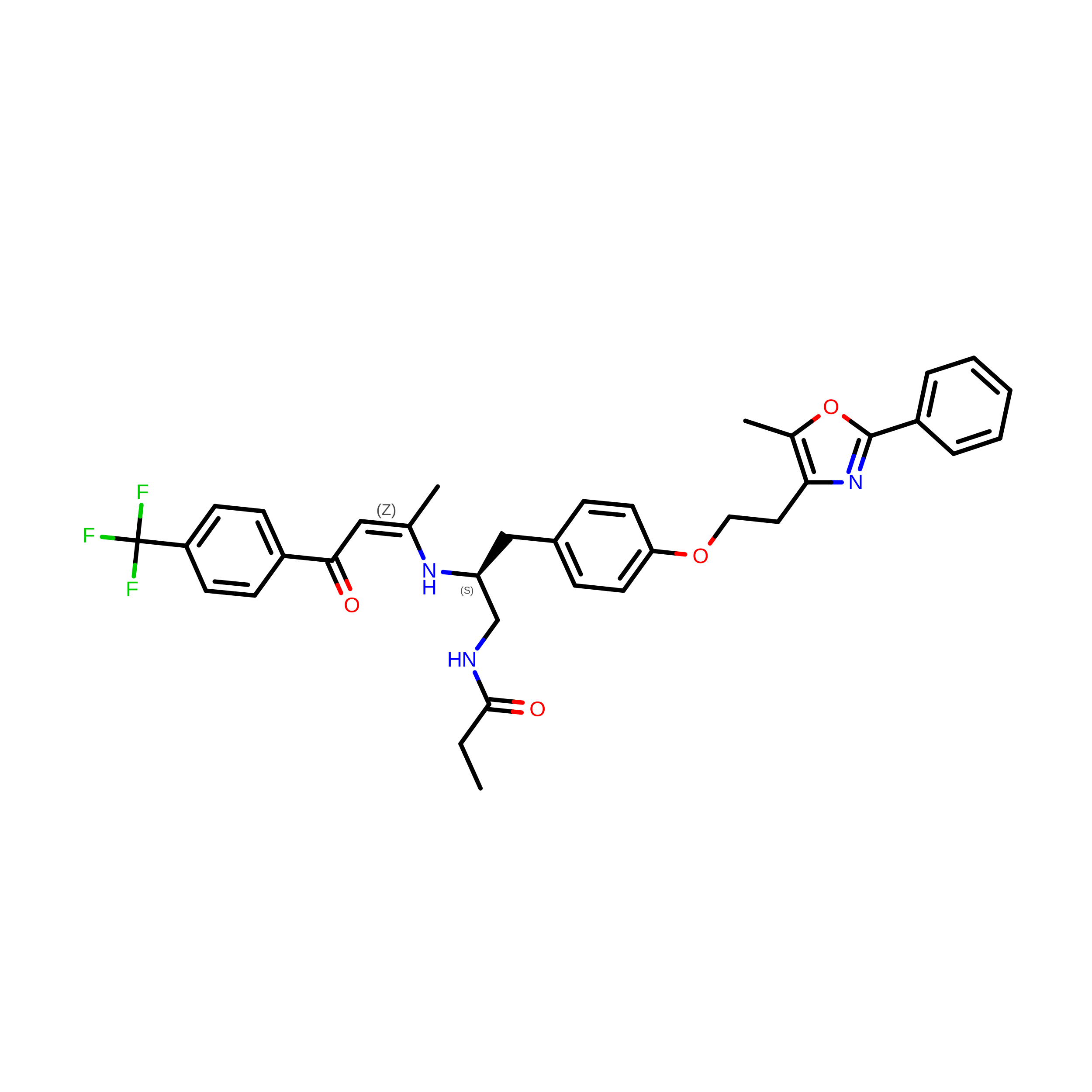
N-((2S)-2-({(1Z)-1-METHYL-3-OXO-3-[4-(TRIFLUOROMETHYL) PHENYL]PROP-1-ENYL}AMINO)-3-{4-[2-(5-METHYL-2-PHENYL-1,3-OXAZOL-4-YL)ETHOXY]PHENYL}PROPYL)PROPANAMIDE
C35 H36 F3 N3 O4
TYEFSRMOUXWTDN-DYQICHDWSA-N

Experimental Data & Validation

Experimental Data

  • Method: X-RAY DIFFRACTION
  • Resolution: 3.00 Å
  • R-Value Free:  0.290 (Depositor) 
  • R-Value Work:  0.258 (Depositor) 
  • R-Value Observed: 0.290 (Depositor) 
Space Group: P 21 21 21
Unit Cell:
Length ( Å )Angle ( ˚ )
a = 103.409α = 90
b = 112.773β = 90
c = 123.954γ = 90
Software Package:
Software NamePurpose
AMoREphasing
CNXrefinement
MAR345data collection
SCALEPACKdata scaling

Structure Validation

View Full Validation Report


Entry History 

Deposition Data

Revision History  (Full details and data files)

  • Version 1.0: 2002-02-20
    Type: Initial release
  • Version 1.1: 2008-04-27
    Changes: Version format compliance
  • Version 1.2: 2011-07-13
    Changes: Version format compliance
  • Version 1.3: 2013-09-18
    Changes: Derived calculations
  • Version 1.4: 2017-02-01
    Changes: Structure summary
  • Version 1.5: 2017-10-11
    Changes: Refinement description
  • Version 1.6: 2023-08-16
    Changes: Data collection, Database references, Derived calculations, Refinement description