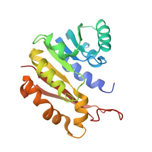
Crystal structure of guanidinoacetate methyltransferase from rat liver: a model structure of protein arginine methyltransferase.
Komoto, J., Huang, Y., Takata, Y., Yamada, T., Konishi, K., Ogawa, H., Gomi, T., Fujioka, M., Takusagawa, F.(2002) J Mol Biology 320: 223-235
- PubMed: 12079381
- DOI: https://doi.org/10.1016/S0022-2836(02)00448-5
- Primary Citation of Related Structures:
1KHH - PubMed Abstract:
Guanidinoacetate methyltransferase (GAMT) is the enzyme that catalyzes the last step of creatine biosynthesis. The enzyme is found in abundance in the livers of all vertebrates. Recombinant rat liver GAMT has been crystallized with S-adenosylhomocysteine (SAH), and the crystal structure has been determined at 2.5 A resolution. The 36 amino acid residues at the N terminus were cleaved during the purification and the truncated enzyme was crystallized. The truncated enzyme forms a dimer, and each subunit contains one SAH molecule in the active site. Arg220 of the partner subunit forms a pair of hydrogen bonds with Asp134 at the guanidinoacetate-binding site. On the basis of the crystal structure, site-directed mutagenesis on Asp134, and chemical modification and limited proteolysis studies, we propose a catalytic mechanism of this enzyme. The truncated GAMT dimer structure can be seen as a ternary complex of protein arginine methyltransferase (one subunit) complexed with a protein substrate (the partner subunit) and the product SAH. Therefore, this structure provides insight into the structure and catalysis of protein arginine methyltransferases.
- Department of Molecular Biosciences, The University of Kansas, 1200 Sunnyside Avenue, 2034 Howorth Hall, Lawrence, KS 66045-7534, USA.
Organizational Affiliation:


