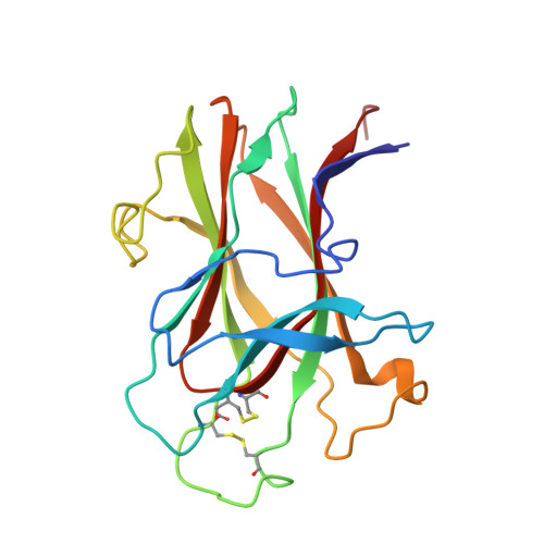
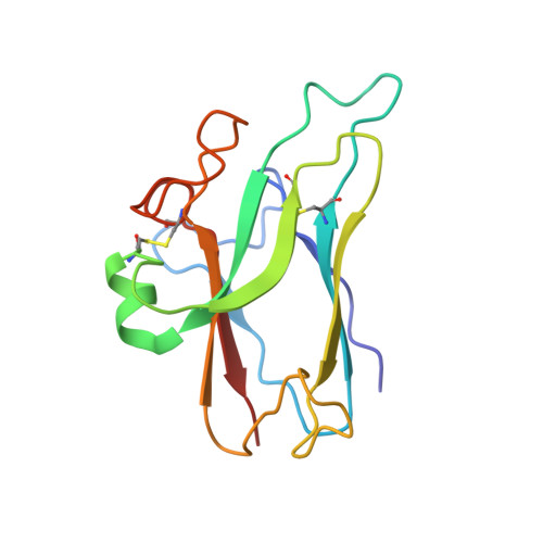
1KGY

Crystal Structure of the EphB2-ephrinB2 complex


Experimental Data Snapshot

  • Method: X-RAY DIFFRACTION
  • Resolution: 2.70 Å
  • R-Value Free: 0.296 
  • R-Value Work: 0.243 

wwPDB Validation   3D Report Full Report


This is version 1.4 of the entry. See complete history


Literature

Crystal structure of an Eph receptor-ephrin complex.

Himanen, J.P.Rajashankar, K.R.Lackmann, M.Cowan, C.A.Henkemeyer, M.Nikolov, D.B.

(2001) Nature 414: 933-938

  • DOI: https://doi.org/10.1038/414933a
  • Primary Citation of Related Structures:  
    1KGY

  • PubMed Abstract: 

    The Eph family of receptor tyrosine kinases and their membrane-anchored ephrin ligands are important in regulating cell-cell interactions as they initiate a unique bidirectional signal transduction cascade whereby information is communicated into both the Eph-expressing and the ephrin-expressing cells. Initially identified as regulators of axon pathfinding and neuronal cell migration, Ephs and ephrins are now known to have roles in many other cell-cell interactions, including those of vascular endothelial cells and specialized epithelia. Here we report the crystal structure of the complex formed between EphB2 and ephrin-B2, determined at 2.7 A resolution. Each Eph receptor binds an ephrin ligand through an expansive dimerization interface dominated by the insertion of an extended ephrin loop into a channel at the surface of the receptor. Two Eph-Ephrin dimers then join to form a tetramer, in which each ligand interacts with two receptors and each receptor interacts with two ligands. The Eph and ephrin molecules are precisely positioned and orientated in these complexes, promoting higher-order clustering and the initiation of bidirectional signalling.


  • Organizational Affiliation

    Cellular Biochemistry and Biophysics Program, Memorial Sloan-Kettering Cancer Center, 1275 York Avenue, New York, New York 10021, USA.


Macromolecules
Find similar proteins by:  (by identity cutoff)  |  3D Structure
Entity ID: 1
MoleculeChains Sequence LengthOrganismDetailsImage
EPHRIN TYPE-B RECEPTOR 2
A, B, C, D
181Mus musculusMutation(s): 0 
EC: 2.7.1.112 (PDB Primary Data), 2.7.10.1 (UniProt)
UniProt & NIH Common Fund Data Resources
Find proteins for P54763 (Mus musculus)
Explore P54763 
Go to UniProtKB:  P54763
IMPC:  MGI:99611
Entity Groups  
Sequence Clusters30% Identity50% Identity70% Identity90% Identity95% Identity100% Identity
UniProt GroupP54763
Sequence Annotations
Expand
  • Reference Sequence
Find similar proteins by:  (by identity cutoff)  |  3D Structure
Entity ID: 2
MoleculeChains Sequence LengthOrganismDetailsImage
EPHRIN-B2
E, F, G, H
138Mus musculusMutation(s): 0 
UniProt
Find proteins for P52800 (Mus musculus)
Explore P52800 
Go to UniProtKB:  P52800
Entity Groups  
Sequence Clusters30% Identity50% Identity70% Identity90% Identity95% Identity100% Identity
UniProt GroupP52800
Sequence Annotations
Expand
  • Reference Sequence
Experimental Data & Validation

Experimental Data

  • Method: X-RAY DIFFRACTION
  • Resolution: 2.70 Å
  • R-Value Free: 0.296 
  • R-Value Work: 0.243 
  • Space Group: P 1
Unit Cell:
Length ( Å )Angle ( ˚ )
a = 78α = 69
b = 78β = 75
c = 78γ = 69
Software Package:
Software NamePurpose
MAR345data collection
SCALEPACKdata scaling
MLPHAREphasing
CNSrefinement

Structure Validation

View Full Validation Report


Entry History 

Deposition Data

Revision History  (Full details and data files)

  • Version 1.0: 2002-05-28
    Type: Initial release
  • Version 1.1: 2008-04-27
    Changes: Version format compliance
  • Version 1.2: 2011-07-13
    Changes: Version format compliance
  • Version 1.3: 2017-02-08
    Changes: Structure summary
  • Version 1.4: 2024-10-30
    Changes: Data collection, Database references, Refinement description, Structure summary