1KFA | pdb_00001kfa

Crystal structure of Fab fragment complexed with gibberellin A4


Experimental Data Snapshot

  • Method: X-RAY DIFFRACTION
  • Resolution: 2.80 Å
  • R-Value Free: 
    0.313 (Depositor), 0.212 (DCC) 
  • R-Value Work: 
    0.235 (Depositor), 0.222 (DCC) 
  • R-Value Observed: 
    0.261 (Depositor) 

Starting Model: experimental
View more details

wwPDB Validation 3D Report Full Report

Validation slider image for 1KFA

This is version 1.5 of the entry. See complete history

Literature

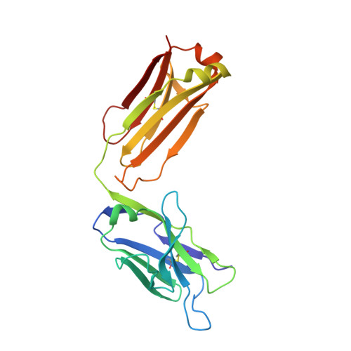
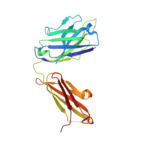
Crystal structure of the liganded anti-gibberellin A(4) antibody 4-B8(8)/E9 Fab fragment.

Murata, T.Fushinobu, S.Nakajima, M.Asami, O.Sassa, T.Wakagi, T.Yamaguchi, I.

(2002) Biochem Biophys Res Commun 293: 489-496

  • DOI: https://doi.org/10.1016/S0006-291X(02)00225-5
  • Primary Citation Related Structures: 
    1KFA

  • PubMed Abstract: 

    Gibberellins, a class of plant hormones, consist of more than 120 members. Only a few of them are recognized by a receptor that remains unknown. The haptenic mouse monoclonal antibody, 4-B8(8)/E9, was generated against gibberellin A(4) (GA(4)) to recognize biologically active GA selectivity, and we attempted to confirm the binding properties between the antibody and GA(4). We carried out an X-ray crystallographic analysis of the 4-B8(8)/E9 Fab fragment complexed with GA(4) at a 2.8 A resolution by using the molecular replacement method. The crystal structure of the Fab fragment showed the typical immunoglobulin fold of the beta-barrel structure which is the common motif of all antibodies. A small hapten-combining site was made up of three heavy chain CDR loops. On the other hand, CDRs of the light chain did not interact directly with GA(4). The C/D rings of the GA(4) molecule were in van der Waals contact mainly with the aromatic side chain of Tyr100AH and Phe100BH of CDR-H3. The 3 beta-hydroxyl and 6 beta-carboxyl groups were, respectively, hydrogen-bonded to the main chain of Ala33H and to the Thr53H heavy chain.


  • Organizational Affiliation
    • Department of Applied Biological Chemistry, Division of Agriculture and Agricultural Life Sciences, The University of Tokyo, 1-1-1 Yayoi, Bunkyo-ku, Tokyo 113-8657, Japan. tks@pgr1.ch.a.u-tokyo.ac.jp

Macromolecule Content 

  • Total Structure Weight: 95.7 kDa 
  • Atom Count: 6,677 
  • Modeled Residue Count: 852 
  • Deposited Residue Count: 876 
  • Unique protein chains: 2

Macromolecules

Find similar proteins by:|  3D Structure
Entity ID: 1
MoleculeChains  Sequence LengthOrganismDetailsImage
monoclonal antibody light chainA [auth L],
C [auth M]
217Mus musculusMutation(s): 0 
Find similar proteins by:|  3D Structure
Entity ID: 2
MoleculeChains  Sequence LengthOrganismDetailsImage
monoclonal antibody heavy chainB [auth H],
D [auth I]
221Mus musculusMutation(s): 0 

Experimental Data & Validation

Experimental Data

  • Method: X-RAY DIFFRACTION
  • Resolution: 2.80 Å
  • R-Value Free:  0.313 (Depositor), 0.212 (DCC) 
  • R-Value Work:  0.235 (Depositor), 0.222 (DCC) 
  • R-Value Observed: 0.261 (Depositor) 
Space Group: C 2 2 21
Unit Cell:
Length ( Å )Angle ( ˚ )
a = 118.076α = 90
b = 150.146β = 90
c = 106.87γ = 90
Software Package:
Software NamePurpose
MOSFLMdata reduction
SCALAdata scaling
CNSrefinement
CCP4data scaling
CNSphasing

Structure Validation

View Full Validation Report


Entry History 

Deposition Data

Revision History  (Full details and data files)

  • Version 1.0: 2002-09-11
    Type: Initial release
  • Version 1.1: 2008-04-27
    Changes: Version format compliance
  • Version 1.2: 2011-07-13
    Changes: Version format compliance
  • Version 1.3: 2013-09-25
    Changes: Source and taxonomy
  • Version 1.4: 2023-10-25
    Changes: Data collection, Database references, Derived calculations, Refinement description
  • Version 1.5: 2024-10-23
    Changes: Structure summary