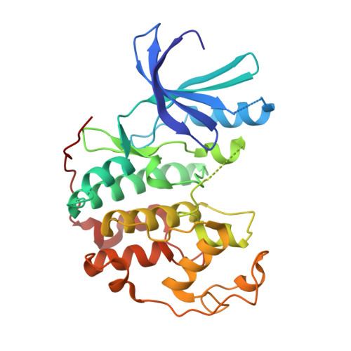
1KE5

CDK2 complexed with N-methyl-4-{[(2-oxo-1,2-dihydro-3H-indol-3-ylidene)methyl]amino}benzenesulfonamide


Experimental Data Snapshot

  • Method: X-RAY DIFFRACTION
  • Resolution: 2.20 Å
  • R-Value Free: 0.224 
  • R-Value Work: 0.184 

wwPDB Validation   3D Report Full Report


Ligand Structure Quality Assessment 


This is version 1.4 of the entry. See complete history


Literature

Oxindole-based inhibitors of cyclin-dependent kinase 2 (CDK2): design, synthesis, enzymatic activities, and X-ray crystallographic analysis.

Bramson, H.N.Corona, J.Davis, S.T.Dickerson, S.H.Edelstein, M.Frye, S.V.Gampe Jr., R.T.Harris, P.A.Hassell, A.Holmes, W.D.Hunter, R.N.Lackey, K.E.Lovejoy, B.Luzzio, M.J.Montana, V.Rocque, W.J.Rusnak, D.Shewchuk, L.Veal, J.M.Walker, D.H.Kuyper, L.F.

(2001) J Med Chem 44: 4339-4358

  • DOI: https://doi.org/10.1021/jm010117d
  • Primary Citation of Related Structures:  
    1KE5, 1KE6, 1KE7, 1KE8, 1KE9

  • PubMed Abstract: 

    Two closely related classes of oxindole-based compounds, 1H-indole-2,3-dione 3-phenylhydrazones and 3-(anilinomethylene)-1,3-dihydro-2H-indol-2-ones, were shown to potently inhibit cyclin-dependent kinase 2 (CDK2). The initial lead compound was prepared as a homologue of the 3-benzylidene-1,3-dihydro-2H-indol-2-one class of kinase inhibitor. Crystallographic analysis of the lead compound bound to CDK2 provided the basis for analogue design. A semiautomated method of ligand docking was used to select compounds for synthesis, and a number of compounds with low nanomolar inhibitory activity versus CDK2 were identified. Enzyme binding determinants for several analogues were evaluated by X-ray crystallography. Compounds in this series inhibited CDK2 with a potency approximately 10-fold greater than that for CDK1. Members of this class of inhibitor cause an arrest of the cell cycle and have shown potential utility in the prevention of chemotherapy-induced alopecia.


  • Organizational Affiliation

    GlaxoSmithKline Inc., Five Moore Drive, Research Triangle Park, North Carolina 27709, USA.


Macromolecules
Find similar proteins by:  (by identity cutoff)  |  3D Structure
Entity ID: 1
MoleculeChains Sequence LengthOrganismDetailsImage
Cell division protein kinase 2298Homo sapiensMutation(s): 0 
EC: 2.7.1.37 (PDB Primary Data), 2.7.11.22 (UniProt)
UniProt & NIH Common Fund Data Resources
Find proteins for P24941 (Homo sapiens)
Explore P24941 
Go to UniProtKB:  P24941
PHAROS:  P24941
GTEx:  ENSG00000123374 
Entity Groups  
Sequence Clusters30% Identity50% Identity70% Identity90% Identity95% Identity100% Identity
UniProt GroupP24941
Sequence Annotations
Expand
  • Reference Sequence
Small Molecules
Ligands 1 Unique
IDChains Name / Formula / InChI Key2D Diagram3D Interactions
LS1
Query on LS1

Download Ideal Coordinates CCD File 
B [auth A]N-METHYL-4-{[(2-OXO-1,2-DIHYDRO-3H-INDOL-3-YLIDENE)METHYL]AMINO}BENZENESULFONAMIDE
C16 H15 N3 O3 S
IKASAFLVQIJQOK-UVTDQMKNSA-N
Binding Affinity Annotations 
IDSourceBinding Affinity
LS1 PDBBind:  1KE5 IC50: 560 (nM) from 1 assay(s)
BindingDB:  1KE5 IC50: 560 (nM) from 1 assay(s)
Experimental Data & Validation

Experimental Data

  • Method: X-RAY DIFFRACTION
  • Resolution: 2.20 Å
  • R-Value Free: 0.224 
  • R-Value Work: 0.184 
  • Space Group: P 21 21 21
Unit Cell:
Length ( Å )Angle ( ˚ )
a = 72.39α = 90
b = 73.96β = 90
c = 54.06γ = 90
Software Package:
Software NamePurpose
X-PLORmodel building
X-PLORrefinement
SCALEPACKdata scaling
X-PLORphasing

Structure Validation

View Full Validation Report


Ligand Structure Quality Assessment 


Entry History 

Deposition Data

Revision History  (Full details and data files)

  • Version 1.0: 2002-05-14
    Type: Initial release
  • Version 1.1: 2008-04-27
    Changes: Version format compliance
  • Version 1.2: 2011-07-13
    Changes: Version format compliance
  • Version 1.3: 2018-01-31
    Changes: Experimental preparation, Refinement description
  • Version 1.4: 2024-02-07
    Changes: Data collection, Database references, Derived calculations