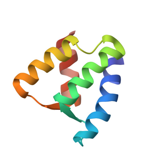
The EF-hand domain: a globally cooperative structural unit.
Nelson, M.R., Thulin, E., Fagan, P.A., Forsen, S., Chazin, W.J.(2002) Protein Sci 11: 198-205
- PubMed: 11790829 Search on PubMedSearch on PubMed Central
- DOI: https://doi.org/10.1110/ps.33302
- Primary Citation Related Structures:
1KCY - PubMed Abstract:
EF-hand Ca(2+)-binding proteins participate in both modulation of Ca(2+) signals and direct transduction of the ionic signal into downstream biochemical events. The range of biochemical functions of these proteins is correlated with differences in the way in which they respond to the binding of Ca(2+). The EF-hand domains of calbindin D(9k) and calmodulin are homologous, yet they respond to the binding of calcium ions in a drastically different manner. A series of comparative analyses of their structures enabled the development of hypotheses about which residues in these proteins control the calcium-induced changes in conformation. To test our understanding of the relationship between protein sequence and structure, we specifically designed the F36G mutation of the EF-hand protein calbindin D(9k) to alter the packing of helices I and II in the apoprotein. The three-dimensional structure of apo F36G was determined in solution by nuclear magnetic resonance spectroscopy and showed that the design was successful. Surprisingly, significant structural perturbations also were found to extend far from the site of mutation. The observation of such long-range effects provides clear evidence that four-helix EF-hand domains should be treated as a single globally cooperative unit. A hypothetical mechanism for how the long-range effects are transmitted is described. Our results support the concept of energetic and structural coupling of the key residues that are crucial for a protein's fold and function.
- Department of Molecular Biology, The Scripps Research Institute, La Jolla, California 92037, USA.
Organizational Affiliation: