1KB4

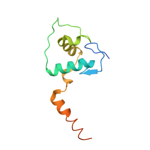
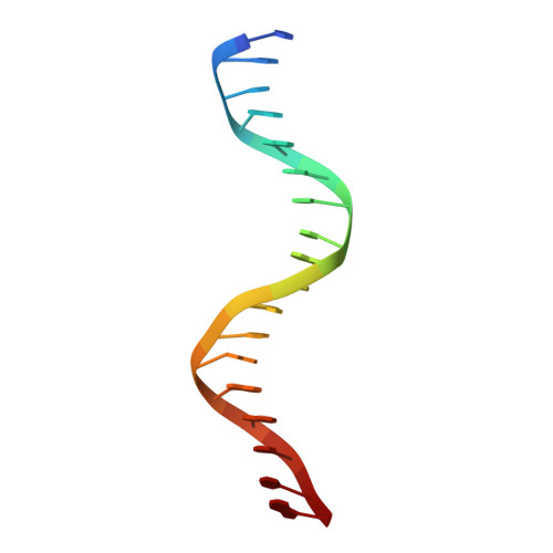
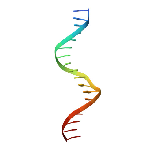
Crystal Structure of VDR DNA-binding Domain Bound to a Canonical Direct Repeat with Three Base Pair Spacer (DR3) Response Element


Experimental Data Snapshot

  • Method: X-RAY DIFFRACTION
  • Resolution: 2.80 Å
  • R-Value Free: 0.272 
  • R-Value Work: 0.214 

Starting Model: experimental
View more details

wwPDB Validation   3D Report Full Report


This is version 1.3 of the entry. See complete history


Literature

Structural basis of VDR-DNA interactions on direct repeat response elements.

Shaffer, P.L.Gewirth, D.T.

(2002) EMBO J 21: 2242-2252

  • DOI: https://doi.org/10.1093/emboj/21.9.2242
  • Primary Citation of Related Structures:  
    1KB2, 1KB4, 1KB6

  • PubMed Abstract: 

    The vitamin D receptor (VDR) forms homo- or heterodimers on response elements composed of two hexameric half-sites separated by 3 bp of spacer DNA. We describe here the crystal structures at 2.7-2.8 A resolution of the VDR DNA-binding region (DBD) in complex with response elements from three different promoters: osteopontin (SPP), canonical DR3 and osteocalcin (OC). These structures reveal the chemical basis for the increased affinity of VDR for the SPP response element, and for the poor stability of the VDR-OC complex, relative to the canonical DR3 response element. The homodimeric protein-protein interface is stabilized by van der Waals interactions and is predominantly non-polar. An extensive alpha-helix at the C-terminal end of the VDR DBD resembles that found in the thyroid hormone receptor (TR), and suggests a mechanism by which VDR and TR discriminate among response elements. Selective structure-based mutations in the asymmetric homodimeric interface result in a VDR DBD protein that is defective in homodimerization but now forms heterodimers with the 9-cis retinoic acid receptor (RXR) DBD.


  • Organizational Affiliation

    Department of Biochemistry, Duke University Medical Center, Durham, NC 27710, USA.


Macromolecules

Find similar proteins by:  (by identity cutoff)  |  3D Structure
Entity ID: 3
MoleculeChains Sequence LengthOrganismDetailsImage
Vitamin D3 ReceptorC [auth A],
D [auth B]
110Homo sapiensMutation(s): 0 
Gene Names: VDR
UniProt & NIH Common Fund Data Resources
Find proteins for P11473 (Homo sapiens)
Explore P11473 
Go to UniProtKB:  P11473
PHAROS:  P11473
GTEx:  ENSG00000111424 
Entity Groups  
Sequence Clusters30% Identity50% Identity70% Identity90% Identity95% Identity100% Identity
UniProt GroupP11473
Sequence Annotations
Expand
  • Reference Sequence

Find similar nucleic acids by:  Sequence   |   3D Structure  

Entity ID: 1
MoleculeChains LengthOrganismImage
5'-D(*CP*AP*CP*AP*GP*GP*TP*CP*AP*CP*GP*AP*AP*GP*GP*TP*CP*A)-3'A [auth C]18N/A
Sequence Annotations
Expand
  • Reference Sequence

Find similar nucleic acids by:  Sequence   |   3D Structure  

Entity ID: 2
MoleculeChains LengthOrganismImage
5'-D(*TP*GP*AP*CP*CP*TP*TP*CP*GP*TP*GP*AP*CP*CP*TP*GP*TP*G)-3'B [auth D]18N/A
Sequence Annotations
Expand
  • Reference Sequence
Experimental Data & Validation

Experimental Data

  • Method: X-RAY DIFFRACTION
  • Resolution: 2.80 Å
  • R-Value Free: 0.272 
  • R-Value Work: 0.214 
  • Space Group: P 43 21 2
Unit Cell:
Length ( Å )Angle ( ˚ )
a = 61.5α = 90
b = 61.5β = 90
c = 241.97γ = 90
Software Package:
Software NamePurpose
CNSrefinement
HKL-2000data reduction
HKL-2000data scaling
CNSphasing

Structure Validation

View Full Validation Report


Entry History 

Deposition Data

Revision History  (Full details and data files)

  • Version 1.0: 2002-05-03
    Type: Initial release
  • Version 1.1: 2008-04-27
    Changes: Version format compliance
  • Version 1.2: 2011-07-13
    Changes: Version format compliance
  • Version 1.3: 2023-08-16
    Changes: Data collection, Database references, Derived calculations, Refinement description