1K02

Crystal Structure of Old Yellow Enzyme Mutant Gln114Asn


Experimental Data Snapshot

  • Method: X-RAY DIFFRACTION
  • Resolution: 2.70 Å
  • R-Value Work: 0.143 
  • R-Value Observed: 0.143 

wwPDB Validation   3D Report Full Report


Ligand Structure Quality Assessment 


This is version 1.4 of the entry. See complete history


Literature

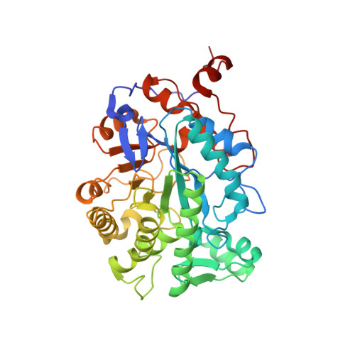
The role of glutamine 114 in old yellow enzyme

Brown, B.J.Hyun, J.Duvvuri, S.D.Karplus, P.A.Massey, V.

(2002) J Biol Chem 277: 2138-2145

  • DOI: https://doi.org/10.1074/jbc.M108453200
  • Primary Citation of Related Structures:  
    1K02, 1K03

  • PubMed Abstract: 

    Glutamine 114 of OYE1 is a well conserved residue in the active site of the Old Yellow Enzyme family. It forms hydrogen bonds to the O2 and N3 of the flavoprotein prosthetic group, FMN. Glutamine 114 was mutated to asparagine, introducing an R-group that is one methylene group shorter. The resultant enzyme was characterized to determine the effect of the mutation on the mechanistic behavior of the enzyme, and the crystal structure was solved to determine the effect of the mutation on the structure of the protein. The Q114N mutation results in little change in the protein structure, moving the amide group of residue 114 out of H-bonding distance, allowing repositioning of the FMN prosthetic group to form new interactions that replace the lost H-bonds. The mutation decreases the ability to bind ligands, as all dissociation constants for substituted phenols are larger than for the wild type enzyme. The rate constant for the reductive half-reaction with beta-NADPH is slightly greater, whereas that for the oxidative half-reaction with 2-cyclohexenone is smaller than for the wild type enzyme. Oxidation with molecular oxygen is biphasic and involves formation and reaction with O(2), a phenomenon that is more pronounced with this mutation than with wild type enzyme. When superoxide dismutase is added to the reaction, we observe a single-phase reaction typical of the wild type enzyme. Turnover reactions using beta-NADPH with 2-cyclohexenone and molecular oxygen were studied to further characterize the mutant enzyme.


  • Organizational Affiliation

    Department of Biological Chemistry, University of Michigan, Ann Arbor, Michigan 48109-0606, USA.


Macromolecules
Find similar proteins by:  (by identity cutoff)  |  3D Structure
Entity ID: 1
MoleculeChains Sequence LengthOrganismDetailsImage
NADPH DEHYDROGENASE 1399Saccharomyces cerevisiaeMutation(s): 1 
EC: 1.6.99.1
UniProt
Find proteins for Q02899 (Saccharomyces pastorianus)
Explore Q02899 
Go to UniProtKB:  Q02899
Entity Groups  
Sequence Clusters30% Identity50% Identity70% Identity90% Identity95% Identity100% Identity
UniProt GroupQ02899
Sequence Annotations
Expand
  • Reference Sequence
Small Molecules
Ligands 2 Unique
IDChains Name / Formula / InChI Key2D Diagram3D Interactions
FMN
Query on FMN

Download Ideal Coordinates CCD File 
C [auth A]FLAVIN MONONUCLEOTIDE
C17 H21 N4 O9 P
FVTCRASFADXXNN-SCRDCRAPSA-N
MG
Query on MG

Download Ideal Coordinates CCD File 
B [auth A]MAGNESIUM ION
Mg
JLVVSXFLKOJNIY-UHFFFAOYSA-N
Experimental Data & Validation

Experimental Data

  • Method: X-RAY DIFFRACTION
  • Resolution: 2.70 Å
  • R-Value Work: 0.143 
  • R-Value Observed: 0.143 
  • Space Group: P 43 21 2
Unit Cell:
Length ( Å )Angle ( ˚ )
a = 142.455α = 90
b = 142.455β = 90
c = 42.928γ = 90
Software Package:
Software NamePurpose
DENZOdata reduction
SCALEPACKdata scaling
CNSrefinement
CNSphasing

Structure Validation

View Full Validation Report


Ligand Structure Quality Assessment 


Entry History 

Deposition Data

Revision History  (Full details and data files)

  • Version 1.0: 2001-09-26
    Type: Initial release
  • Version 1.1: 2008-04-27
    Changes: Version format compliance
  • Version 1.2: 2011-07-13
    Changes: Version format compliance
  • Version 1.3: 2021-10-27
    Changes: Database references, Derived calculations
  • Version 1.4: 2024-02-07
    Changes: Data collection