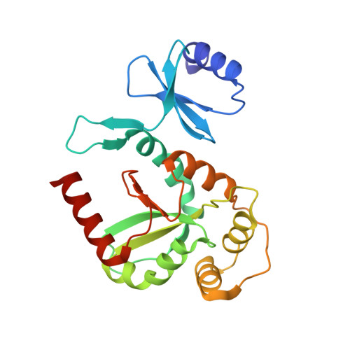
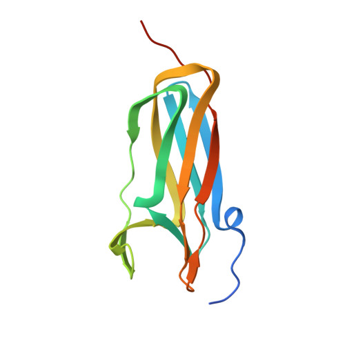
1JZD

DsbC-DsbDalpha complex


Experimental Data Snapshot

  • Method: X-RAY DIFFRACTION
  • Resolution: 2.30 Å
  • R-Value Free: 0.293 
  • R-Value Work: 0.234 

Starting Model: experimental
View more details

wwPDB Validation   3D Report Full Report


This is version 1.4 of the entry. See complete history


Literature

The Disulfide Bond Isomerase DsbC is Activated by an Immunoglobulin-fold Thiol Oxidoreductase: Crystal structure of the DsbC-DsbDalpha complex.

Haebel, P.W.Goldstone, D.Katzen, F.Beckwith, J.Metcalf, P.

(2002) EMBO J 21: 4774-4784

  • DOI: https://doi.org/10.1093/emboj/cdf489
  • Primary Citation of Related Structures:  
    1JPE, 1JZD, 1JZO

  • PubMed Abstract: 

    The Escherichia coli disulfide bond isomerase DsbC rearranges incorrect disulfide bonds during oxidative protein folding. It is specifically activated by the periplasmic N-terminal domain (DsbDalpha) of the transmembrane electron transporter DsbD. An intermediate of the electron transport reaction was trapped, yielding a covalent DsbC-DsbDalpha complex. The 2.3 A crystal structure of the complex shows for the first time the specific interactions between two thiol oxidoreductases. DsbDalpha is a novel thiol oxidoreductase with the active site cysteines embedded in an immunoglobulin fold. It binds into the central cleft of the V-shaped DsbC dimer, which assumes a closed conformation on complex formation. Comparison of the complex with oxidized DsbDalpha reveals major conformational changes in a cap structure that regulates the accessibility of the DsbDalpha active site. Our results explain how DsbC is selectively activated by DsbD using electrons derived from the cytoplasm.


  • Organizational Affiliation

    School of Biological Sciences, University of Auckland, Private Bag 92019, Auckland, New Zealandand.


Macromolecules
Find similar proteins by:  (by identity cutoff)  |  3D Structure
Entity ID: 1
MoleculeChains Sequence LengthOrganismDetailsImage
thiol:disulfide interchange protein dsbc
A, B
220Escherichia coliMutation(s): 1 
Gene Names: dsbC
UniProt
Find proteins for P0AEG6 (Escherichia coli (strain K12))
Explore P0AEG6 
Go to UniProtKB:  P0AEG6
Entity Groups  
Sequence Clusters30% Identity50% Identity70% Identity90% Identity95% Identity100% Identity
UniProt GroupP0AEG6
Sequence Annotations
Expand
  • Reference Sequence
Find similar proteins by:  (by identity cutoff)  |  3D Structure
Entity ID: 2
MoleculeChains Sequence LengthOrganismDetailsImage
thiol:disulfide interchange protein dsbd132Escherichia coliMutation(s): 1 
Gene Names: dsbD
UniProt
Find proteins for P36655 (Escherichia coli (strain K12))
Explore P36655 
Go to UniProtKB:  P36655
Entity Groups  
Sequence Clusters30% Identity50% Identity70% Identity90% Identity95% Identity100% Identity
UniProt GroupP36655
Sequence Annotations
Expand
  • Reference Sequence
Experimental Data & Validation

Experimental Data

  • Method: X-RAY DIFFRACTION
  • Resolution: 2.30 Å
  • R-Value Free: 0.293 
  • R-Value Work: 0.234 
  • Space Group: P 41 21 2
Unit Cell:
Length ( Å )Angle ( ˚ )
a = 68.91α = 90
b = 68.91β = 90
c = 230.336γ = 90
Software Package:
Software NamePurpose
MOSFLMdata reduction
SCALAdata scaling
MOLREPphasing
CNSrefinement
CCP4data scaling

Structure Validation

View Full Validation Report


Entry History 

Deposition Data

Revision History  (Full details and data files)

  • Version 1.0: 2003-03-08
    Type: Initial release
  • Version 1.1: 2008-04-27
    Changes: Version format compliance
  • Version 1.2: 2011-07-13
    Changes: Version format compliance
  • Version 1.3: 2021-10-27
    Changes: Data collection, Database references
  • Version 1.4: 2023-08-16
    Changes: Data collection, Refinement description