1JX6 | pdb_00001jx6

CRYSTAL STRUCTURE OF LUXP FROM VIBRIO HARVEYI COMPLEXED WITH AUTOINDUCER-2


Experimental Data Snapshot

  • Method: X-RAY DIFFRACTION
  • Resolution: 1.50 Å
  • R-Value Free: 
    0.239 (Depositor), 0.234 (DCC) 
  • R-Value Work: 
    0.214 (Depositor), 0.210 (DCC) 
  • R-Value Observed: 
    0.214 (Depositor) 

wwPDB Validation 3D Report Full Report

Validation slider image for 1JX6

Ligand Structure Quality Assessment 


This is version 1.3 of the entry. See complete history

Literature

Structural identification of a bacterial quorum-sensing signal containing boron.

Chen, X.Schauder, S.Potier, N.Van Dorsselaer, A.Pelczer, I.Bassler, B.L.Hughson, F.M.

(2002) Nature 415: 545-549

  • DOI: https://doi.org/10.1038/415545a
  • Primary Citation Related Structures: 
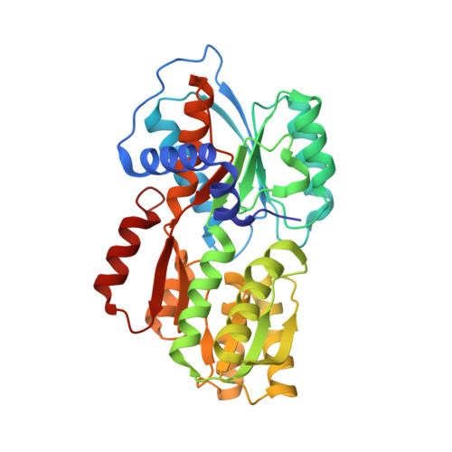
    1JX6

  • PubMed Abstract: 

    Cell-cell communication in bacteria is accomplished through the exchange of extracellular signalling molecules called autoinducers. This process, termed quorum sensing, allows bacterial populations to coordinate gene expression. Community cooperation probably enhances the effectiveness of processes such as bioluminescence, virulence factor expression, antibiotic production and biofilm development. Unlike other autoinducers, which are specific to a particular species of bacteria, a recently discovered autoinducer (AI-2) is produced by a large number of bacterial species. AI-2 has been proposed to serve as a 'universal' signal for inter-species communication. The chemical identity of AI-2 has, however, proved elusive. Here we present the crystal structure of an AI-2 sensor protein, LuxP, in a complex with autoinducer. The bound ligand is a furanosyl borate diester that bears no resemblance to previously characterized autoinducers. Our findings suggest that addition of naturally occurring borate to an AI-2 precursor generates active AI-2. Furthermore, they indicate a potential biological role for boron, an element required by a number of organisms but for unknown reasons.


  • Organizational Affiliation
    • Department of Molecular Biology, Princeton University, Princeton, New Jersey 08544-1014, USA.

Macromolecule Content 

  • Total Structure Weight: 39.3 kDa 
  • Atom Count: 3,073 
  • Modeled Residue Count: 338 
  • Deposited Residue Count: 342 
  • Unique protein chains: 1

Macromolecules

Find similar proteins by:|  3D Structure
Entity ID: 1
MoleculeChains  Sequence LengthOrganismDetailsImage
LUXP PROTEIN342Vibrio harveyiMutation(s): 0 
Gene Names: LUXP
UniProt
Find proteins for P54300 (Vibrio harveyi)
Explore P54300 
Go to UniProtKB:  P54300
Entity Groups
Sequence Clusters30% Identity50% Identity70% Identity90% Identity95% Identity100% Identity
UniProt GroupP54300
Sequence Annotations
Expand
Reference Sequence

Small Molecules

Ligands 2 Unique
IDChains Name / Formula / InChI Key2D Diagram3D Interactions
AI2

Query on AI2


Download:Ideal Coordinates CCD File
C [auth A]3A-METHYL-5,6-DIHYDRO-FURO[2,3-D][1,3,2]DIOXABOROLE-2,2,6,6A-TETRAOL
C5 H10 B O7
ACKRRKSNOOISSG-VPENINKCSA-N
CA

Query on CA


Download:Ideal Coordinates CCD File
B [auth A]CALCIUM ION
Ca
BHPQYMZQTOCNFJ-UHFFFAOYSA-N

Experimental Data & Validation

Experimental Data

  • Method: X-RAY DIFFRACTION
  • Resolution: 1.50 Å
  • R-Value Free:  0.239 (Depositor), 0.234 (DCC) 
  • R-Value Work:  0.214 (Depositor), 0.210 (DCC) 
  • R-Value Observed: 0.214 (Depositor) 
Space Group: P 1 21 1
Unit Cell:
Length ( Å )Angle ( ˚ )
a = 44.268α = 90
b = 77.467β = 96.37
c = 52.008γ = 90
Software Package:
Software NamePurpose
MLPHAREphasing
CNSrefinement
MARMADdata reduction
SCALEPACKdata scaling

Structure Validation

View Full Validation Report


Ligand Structure Quality Assessment 


Entry History 

Deposition Data

Revision History  (Full details and data files)

  • Version 1.0: 2002-02-04
    Type: Initial release
  • Version 1.1: 2008-04-27
    Changes: Version format compliance
  • Version 1.2: 2011-07-13
    Changes: Version format compliance
  • Version 1.3: 2024-02-07
    Changes: Data collection, Database references, Derived calculations