1JUH | pdb_00001juh

Crystal Structure of Quercetin 2,3-dioxygenase


Experimental Data Snapshot

  • Method: X-RAY DIFFRACTION
  • Resolution: 1.60 Å
  • R-Value Free: 
    0.189 (Depositor), 0.204 (DCC) 
  • R-Value Work: 
    0.162 (Depositor), 0.179 (DCC) 
  • R-Value Observed: 
    0.162 (Depositor) 

wwPDB Validation 3D Report Full Report

Validation slider image for 1JUH

Ligand Structure Quality Assessment 


This is version 2.1 of the entry. See complete history

Literature

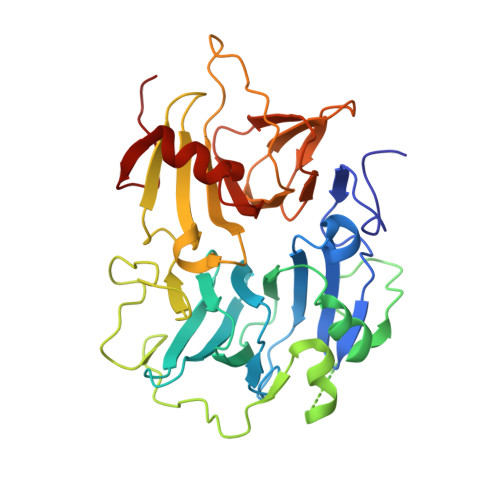
Crystal structure of the copper-containing quercetin 2,3-dioxygenase from Aspergillus japonicus.

Fusetti, F.Schroter, K.H.Steiner, R.A.van Noort, P.I.Pijning, T.Rozeboom, H.J.Kalk, K.H.Egmond, M.R.Dijkstra, B.W.

(2002) Structure 10: 259-268

  • DOI: https://doi.org/10.1016/s0969-2126(02)00704-9
  • Primary Citation Related Structures: 
    1JUH

  • PubMed Abstract: 

    Quercetin 2,3-dioxygenase is a copper-containing enzyme that catalyzes the insertion of molecular oxygen into polyphenolic flavonols. Dioxygenation catalyzed by iron-containing enzymes has been studied extensively, but dioxygenases employing other metal cofactors are poorly understood. We determined the crystal structure of quercetin 2,3-dioxygenase at 1.6 A resolution. The enzyme forms homodimers, which are stabilized by an N-linked heptasaccharide at the dimer interface. The mononuclear type 2 copper center displays two distinct geometries: a distorted tetrahedral coordination, formed by His66, His68, His112, and a water molecule, and a distorted trigonal bipyramidal environment, which additionally comprises Glu73. Manual docking of the substrate quercetin into the active site showed that the different geometries of the copper site might be of catalytic importance.


  • Organizational Affiliation
    • Laboratory of Biophysical Chemistry, Department of Chemistry, University of Groningen, Nijenborgh 4, 9747 AG Groningen, The Netherlands.

Macromolecule Content 

  • Total Structure Weight: 160.11 kDa 
  • Atom Count: 12,719 
  • Modeled Residue Count: 1,337 
  • Deposited Residue Count: 1,400 
  • Unique protein chains: 1

Macromolecules

Find similar proteins by:|  3D Structure
Entity ID: 1
MoleculeChains  Sequence LengthOrganismDetailsImage
quercetin 2,3-dioxygenase
A, B, C, D
350Aspergillus japonicusMutation(s): 0 
EC: 1.13.11.24
UniProt
Find proteins for Q7SIC2 (Aspergillus japonicus)
Explore Q7SIC2 
Go to UniProtKB:  Q7SIC2
Entity Groups
Sequence Clusters30% Identity50% Identity70% Identity90% Identity95% Identity100% Identity
UniProt GroupQ7SIC2
Glycosylation
Glycosylation Sites: 4
Sequence Annotations
Expand
Reference Sequence

Oligosaccharides

Help  
Entity ID: 2
MoleculeChains Length2D Diagram GlycosylationD Interactions
alpha-D-mannopyranose-(1-2)-alpha-D-mannopyranose-(1-3)-[alpha-D-mannopyranose-(1-3)-alpha-D-mannopyranose-(1-6)]beta-D-mannopyranose-(1-4)-2-acetamido-2-deoxy-beta-D-glucopyranose-(1-4)-2-acetamido-2-deoxy-beta-D-glucopyranose
E
7N-Glycosylation
Glycosylation Resources
GlyTouCan: G07617FP
GlyCosmos: G07617FP
GlyGen: G07617FP
Entity ID: 3
MoleculeChains Length2D Diagram GlycosylationD Interactions
alpha-D-mannopyranose-(1-3)-beta-D-mannopyranose-(1-4)-2-acetamido-2-deoxy-beta-D-glucopyranose-(1-4)-2-acetamido-2-deoxy-beta-D-glucopyranose
F
4N-Glycosylation
Glycosylation Resources
GlyTouCan: G81315DD
GlyCosmos: G81315DD
GlyGen: G81315DD
Entity ID: 4
MoleculeChains Length2D Diagram GlycosylationD Interactions
alpha-D-mannopyranose-(1-6)-beta-D-mannopyranose-(1-4)-2-acetamido-2-deoxy-beta-D-glucopyranose-(1-4)-2-acetamido-2-deoxy-beta-D-glucopyranose
G
4N-Glycosylation
Glycosylation Resources
GlyTouCan: G22573RC
GlyCosmos: G22573RC
GlyGen: G22573RC
Entity ID: 5
MoleculeChains Length2D Diagram GlycosylationD Interactions
2-acetamido-2-deoxy-beta-D-glucopyranose-(1-4)-2-acetamido-2-deoxy-beta-D-glucopyranose
H
2N-Glycosylation
Glycosylation Resources
GlyTouCan: G42666HT
GlyCosmos: G42666HT
GlyGen: G42666HT

Small Molecules

Ligands 3 Unique
IDChains Name / Formula / InChI Key2D Diagram3D Interactions
NAG

Query on NAG


Download:Ideal Coordinates CCD File
I [auth A]
IA [auth C]
J [auth A]
JA [auth C]
K [auth A]
I [auth A],
IA [auth C],
J [auth A],
JA [auth C],
K [auth A],
KA [auth C],
L [auth A],
W [auth B],
WA [auth D],
X [auth B],
XA [auth D],
Y [auth B],
YA [auth D]
2-acetamido-2-deoxy-beta-D-glucopyranose
C8 H15 N O6
OVRNDRQMDRJTHS-FMDGEEDCSA-N
CU

Query on CU


Download:Ideal Coordinates CCD File
LA [auth C],
M [auth A],
Z [auth B],
ZA [auth D]
COPPER (II) ION
Cu
JPVYNHNXODAKFH-UHFFFAOYSA-N
EDO

Query on EDO


Download:Ideal Coordinates CCD File
AA [auth B]
AB [auth D]
BA [auth B]
BB [auth D]
CA [auth B]
AA [auth B],
AB [auth D],
BA [auth B],
BB [auth D],
CA [auth B],
CB [auth D],
DA [auth B],
DB [auth D],
EA [auth B],
EB [auth D],
FA [auth B],
GA [auth B],
HA [auth B],
MA [auth C],
N [auth A],
NA [auth C],
O [auth A],
OA [auth C],
P [auth A],
PA [auth C],
Q [auth A],
QA [auth C],
R [auth A],
RA [auth C],
S [auth A],
SA [auth C],
T [auth A],
TA [auth C],
U [auth A],
UA [auth C],
V [auth A],
VA [auth C]
1,2-ETHANEDIOL
C2 H6 O2
LYCAIKOWRPUZTN-UHFFFAOYSA-N

Experimental Data & Validation

Experimental Data

  • Method: X-RAY DIFFRACTION
  • Resolution: 1.60 Å
  • R-Value Free:  0.189 (Depositor), 0.204 (DCC) 
  • R-Value Work:  0.162 (Depositor), 0.179 (DCC) 
  • R-Value Observed: 0.162 (Depositor) 
Space Group: P 1 21 1
Unit Cell:
Length ( Å )Angle ( ˚ )
a = 108.55α = 90
b = 55.78β = 98.31
c = 123.68γ = 90
Software Package:
Software NamePurpose
PHASESphasing
DMmodel building
CNSrefinement
DENZOdata reduction
SCALEPACKdata scaling
DMphasing

Structure Validation

View Full Validation Report


Ligand Structure Quality Assessment 


Entry History 

Deposition Data

Revision History  (Full details and data files)

  • Version 1.0: 2002-05-22
    Type: Initial release
  • Version 1.1: 2008-04-01
    Changes: Version format compliance
  • Version 1.2: 2011-07-13
    Changes: Non-polymer description, Version format compliance
  • Version 1.3: 2018-10-03
    Changes: Data collection
  • Version 1.4: 2020-01-22
    Changes: Advisory, Database references, Derived calculations
  • Version 2.0: 2020-07-29
    Type: Remediation
    Reason: Carbohydrate remediation
    Changes: Atomic model, Data collection, Derived calculations, Structure summary
  • Version 2.1: 2024-11-20
    Changes: Data collection, Database references, Structure summary