1JPN | pdb_00001jpn

GMPPNP Complex of SRP GTPase NG Domain


Experimental Data Snapshot

  • Method: X-RAY DIFFRACTION
  • Resolution: 1.90 Å
  • R-Value Free: 
    0.241 (Depositor), 0.524 (DCC) 
  • R-Value Work: 
    0.190 (Depositor), 0.521 (DCC) 

Starting Model: experimental
View more details

wwPDB Validation 3D Report Full Report

Validation slider image for 1JPN

Ligand Structure Quality Assessment 


This is version 1.3 of the entry. See complete history

Literature

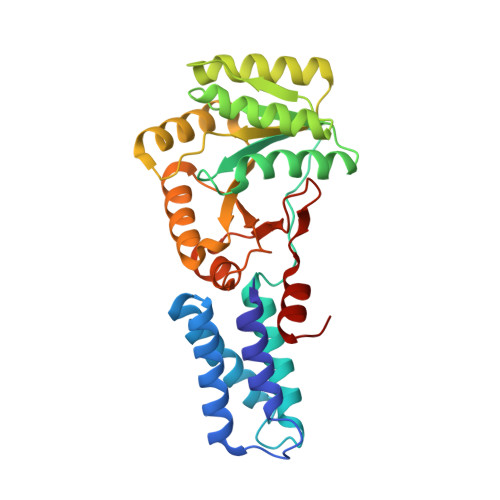
The conformation of bound GMPPNP suggests a mechanism for gating the active site of the SRP GTPase.

Padmanabhan, S.Freymann, D.M.

(2001) Structure 9: 859-867

  • DOI: https://doi.org/10.1016/s0969-2126(01)00641-4
  • Primary Citation Related Structures: 
    1JPJ, 1JPN

  • PubMed Abstract: 

    The signal recognition particle (SRP) is a phylogenetically conserved ribonucleoprotein that mediates cotranslational targeting of secreted and membrane proteins to the membrane. Targeting is regulated by GTP binding and hydrolysis events that require direct interaction between structurally homologous "NG" GTPase domains of the SRP signal recognition subunit and its membrane-associated receptor, SR alpha. Structures of both the apo and GDP bound NG domains of the prokaryotic SRP54 homolog, Ffh, and the prokaryotic receptor homolog, FtsY, have been determined. The structural basis for the GTP-dependent interaction between the two proteins, however, remains unknown. We report here two structures of the NG GTPase of Ffh from Thermus aquaticus bound to the nonhydrolyzable GTP analog GMPPNP. Both structures reveal an unexpected binding mode in which the beta-phosphate is kinked away from the binding site and magnesium is not bound. Binding of the GTP analog in the canonical conformation found in other GTPase structures is precluded by constriction of the phosphate binding P loop. The structural difference between the Ffh complex and other GTPases suggests a specific conformational change that must accompany movement of the nucleotide from an "inactive" to an "active" binding mode. Conserved side chains of the GTPase sequence motifs unique to the SRP subfamily may function to gate formation of the active GTP bound conformation. Exposed hydrophobic residues provide an interaction surface that may allow regulation of the GTP binding conformation, and thus activation of the GTPase, during the association of SRP with its receptor.


  • Organizational Affiliation
    • Department of Molecular Pharmacology and Biological Chemistry, Northwestern University Medical School, Chicago, IL 60611, USA.

Macromolecule Content 

  • Total Structure Weight: 66.31 kDa 
  • Atom Count: 5,117 
  • Modeled Residue Count: 591 
  • Deposited Residue Count: 592 
  • Unique protein chains: 1

Macromolecules

Find similar proteins by:|  3D Structure
Entity ID: 1
MoleculeChains  Sequence LengthOrganismDetailsImage
SIGNAL RECOGNITION PARTICLE PROTEINA [auth B],
B [auth A]
296Thermus aquaticusMutation(s): 0 
EC: 3.6.5.4
UniProt
Find proteins for O07347 (Thermus aquaticus)
Explore O07347 
Go to UniProtKB:  O07347
Entity Groups
Sequence Clusters30% Identity50% Identity70% Identity90% Identity95% Identity100% Identity
UniProt GroupO07347
Sequence Annotations
Expand
Reference Sequence

Small Molecules

Ligands 3 Unique
IDChains Name / Formula / InChI Key2D Diagram3D Interactions
GNP

Query on GNP


Download:Ideal Coordinates CCD File
D [auth B],
H [auth A]
PHOSPHOAMINOPHOSPHONIC ACID-GUANYLATE ESTER
C10 H17 N6 O13 P3
UQABYHGXWYXDTK-UUOKFMHZSA-N
ACY

Query on ACY


Download:Ideal Coordinates CCD File
E [auth B],
F [auth B],
I [auth A],
J [auth A]
ACETIC ACID
C2 H4 O2
QTBSBXVTEAMEQO-UHFFFAOYSA-N
CA

Query on CA


Download:Ideal Coordinates CCD File
C [auth B],
G [auth A]
CALCIUM ION
Ca
BHPQYMZQTOCNFJ-UHFFFAOYSA-N

Experimental Data & Validation

Experimental Data

  • Method: X-RAY DIFFRACTION
  • Resolution: 1.90 Å
  • R-Value Free:  0.241 (Depositor), 0.524 (DCC) 
  • R-Value Work:  0.190 (Depositor), 0.521 (DCC) 
Space Group: C 1 2 1
Unit Cell:
Length ( Å )Angle ( ˚ )
a = 108.815α = 90
b = 54.531β = 97.42
c = 99.078γ = 90
Software Package:
Software NamePurpose
AMoREphasing
X-PLORrefinement
DENZOdata reduction
SCALEPACKdata scaling

Structure Validation

View Full Validation Report


Ligand Structure Quality Assessment 


Entry History 

Deposition Data

Revision History  (Full details and data files)

  • Version 1.0: 2002-02-02
    Type: Initial release
  • Version 1.1: 2008-04-27
    Changes: Version format compliance
  • Version 1.2: 2011-07-13
    Changes: Version format compliance
  • Version 1.3: 2023-08-16
    Changes: Data collection, Database references, Derived calculations, Refinement description