


Crystal 1.57-A Crystal Structure of HI1317
Bonander, N., Tordova, M., Howard, A.J., Eisenstein, E., Gilliland, G.To be published.
Experimental Data Snapshot
wwPDB Validation 3D Report Full Report

Entity ID: 1 | |||||
|---|---|---|---|---|---|
| Molecule | Chains | Sequence Length | Organism | Details | Image |
| HI1317 | 270 | Haemophilus influenzae | Mutation(s): 0 Gene Names: HI1317 EC: 5.1.3.15 | 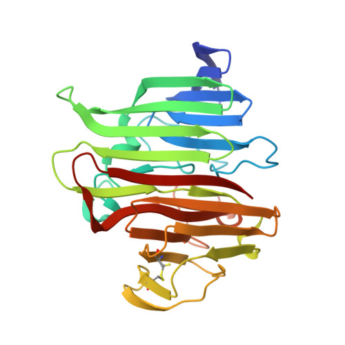 | |
UniProt | |||||
Entity Groups | |||||
| Sequence Clusters | 30% Identity50% Identity70% Identity90% Identity95% Identity100% Identity | ||||
| UniProt Group | Q9RP27 | ||||
Sequence AnnotationsExpand | |||||
Reference Sequence | |||||
| Ligands 2 Unique | |||||
|---|---|---|---|---|---|
| ID | Chains | Name / Formula / InChI Key | 2D Diagram | 3D Interactions | |
| TRS Download:Ideal Coordinates CCD File | C [auth A], D [auth A] | 2-AMINO-2-HYDROXYMETHYL-PROPANE-1,3-DIOL C4 H12 N O3 LENZDBCJOHFCAS-UHFFFAOYSA-O |  | ||
| SO4 Download:Ideal Coordinates CCD File | B [auth A] | SULFATE ION O4 S QAOWNCQODCNURD-UHFFFAOYSA-L |  | ||
| Length ( Å ) | Angle ( ˚ ) |
|---|---|
| a = 80.54 | α = 90 |
| b = 38.86 | β = 110.16 |
| c = 92.36 | γ = 90 |
| Software Name | Purpose |
|---|---|
| HKL-2000 | data reduction |
| REFMAC | refinement |
| HKL-2000 | data scaling |


