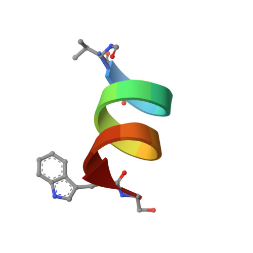
Structures of Gramicidins A, B, and C Incorporated Into Sodium Dodecyl Sulfate Micelles.
Townsley, L.E., Tucker, W.A., Sham, S., Hinton, J.F.(2001) Biochemistry 40: 11676
- PubMed: 11570868
- DOI: https://doi.org/10.1021/bi010942w
- Primary Citation of Related Structures:
1JNO, 1JO3, 1JO4 - PubMed Abstract:
Gramicidins A, B, and C are the three most abundant, naturally occurring analogues of this family of channel-forming antibiotic. GB and GC differ from the parent pentadecapeptide, GA, by single residue mutations, W11F and W11Y, respectively. Although these mutations occur in the cation binding region of the channel, they do not affect monovalent cation specificity, but are known to alter cation-binding affinities, thermodynamic parameters of cation binding, conductance and the activation energy for ion transport. The structures of all three analogues incorporated into deuterated sodium dodecyl sulfate micelles have been obtained using solution state 2D-NMR spectroscopy and molecular modeling. For the first time, a rigorous comparison of the 3D structures of these analogues reveals that the amino acid substitutions do not have a significant effect on backbone conformation, thus eliminating channel differences as the cause of variations in transport properties. Variable positions of methyl groups in valine and leucine residues have been linked to molecular motions and are not likely to affect ion flow through the channel. Thus, it is concluded that changes in the magnitude and orientation of the dipole moment at residue 11 are responsible for altering monovalent cation transport.
- Department of Chemistry and Biochemistry, University of Arkansas, Fayetteville, Arkansas 72701, USA.
Organizational Affiliation:


