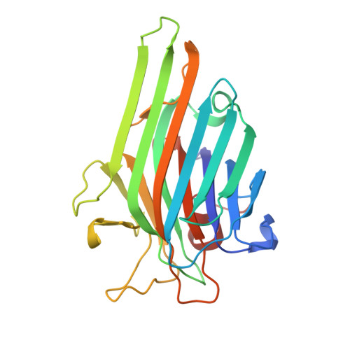
1JN2

Crystal Structure of meso-tetrasulphonatophenyl porphyrin complexed with Concanavalin A


Experimental Data Snapshot

  • Method: X-RAY DIFFRACTION
  • Resolution: 1.90 Å
  • R-Value Free: 0.232 
  • R-Value Work: 0.195 

wwPDB Validation   3D Report Full Report


This is version 1.4 of the entry. See complete history


Literature

Functional equality in the absence of structural similarity: an added dimension to molecular mimicry

Goel, M.Jain, D.Kaur, K.J.Kenoth, R.Maiya, B.G.Swamy, M.J.Salunke, D.M.

(2000) J Biol Chem 276: 39277-39281

  • DOI: https://doi.org/10.1074/jbc.M105387200
  • Primary Citation of Related Structures:  
    1JN2

  • PubMed Abstract: 

    The crystal structure of meso-tetrasulfonatophenylporphyrin complexed with concanavalin A (ConA) was determined at 1.9 A resolution. Comparison of this structure with that of ConA bound to methyl alpha-d-mannopyranoside provided direct structural evidence of molecular mimicry in the context of ligand receptor binding. The sulfonatophenyl group of meso-tetrasulfonatophenylporphyrin occupies the same binding site on ConA as that of methyl alpha-d-mannopyranoside, a natural ligand. A pair of stacked porphyrin molecules stabilizes the crystal structure by end-to-end cross-linking with ConA resulting in a network similar to that observed upon agglutination of cells by lectins. The porphyrin binds to ConA predominantly through hydrogen bonds and water-mediated interactions. The sandwiched water molecules in the complex play a cementing role, facilitating favorable binding of porphyrin. Seven of the eight hydrogen bonds observed between methyl alpha-d-mannopyranoside and ConA are mimicked by the sulfonatophenyl group of porphyrin after incorporating two water molecules. Thus, the similarity in chemical interactions was manifested in terms of functional mimicry despite the obvious structural dissimilarity between the sugar and the porphyrin.


  • Organizational Affiliation

    Structural Biology Unit, National Institute of Immunology, New Delhi 110067, India.


Macromolecules
Find similar proteins by:  (by identity cutoff)  |  3D Structure
Entity ID: 1
MoleculeChains Sequence LengthOrganismDetailsImage
Concanavalin-AA [auth P]237Canavalia ensiformisMutation(s): 0 
UniProt
Find proteins for P02866 (Canavalia ensiformis)
Explore P02866 
Go to UniProtKB:  P02866
Entity Groups  
Sequence Clusters30% Identity50% Identity70% Identity90% Identity95% Identity100% Identity
UniProt GroupP02866
Sequence Annotations
Expand
  • Reference Sequence
Small Molecules
Ligands 3 Unique
IDChains Name / Formula / InChI Key2D Diagram3D Interactions
SFP
Query on SFP

Download Ideal Coordinates CCD File 
D [auth P]5,10,15,20-TETRAKIS(4-SULPFONATOPHENYL)-21H,23H-PORPHINE
C44 H34 N4 O12 S4
OVPFWIRROXFHGQ-LWQDQPMZSA-J
MN
Query on MN

Download Ideal Coordinates CCD File 
C [auth P]MANGANESE (II) ION
Mn
WAEMQWOKJMHJLA-UHFFFAOYSA-N
CA
Query on CA

Download Ideal Coordinates CCD File 
B [auth P]CALCIUM ION
Ca
BHPQYMZQTOCNFJ-UHFFFAOYSA-N
Binding Affinity Annotations 
IDSourceBinding Affinity
SFP PDBBind:  1JN2 Kd: 8.20e+4 (nM) from 1 assay(s)
Experimental Data & Validation

Experimental Data

  • Method: X-RAY DIFFRACTION
  • Resolution: 1.90 Å
  • R-Value Free: 0.232 
  • R-Value Work: 0.195 
  • Space Group: F 2 2 2
Unit Cell:
Length ( Å )Angle ( ˚ )
a = 106α = 90
b = 117.3β = 90
c = 126γ = 90
Software Package:
Software NamePurpose
DENZOdata reduction
SCALEPACKdata scaling
AMoREphasing
CNSrefinement

Structure Validation

View Full Validation Report


Entry History 

Deposition Data

Revision History  (Full details and data files)

  • Version 1.0: 2003-07-01
    Type: Initial release
  • Version 1.1: 2008-04-27
    Changes: Version format compliance
  • Version 1.2: 2011-07-13
    Changes: Derived calculations, Version format compliance
  • Version 1.3: 2017-06-28
    Changes: Database references, Source and taxonomy, Structure summary
  • Version 1.4: 2024-02-07
    Changes: Data collection, Database references, Derived calculations