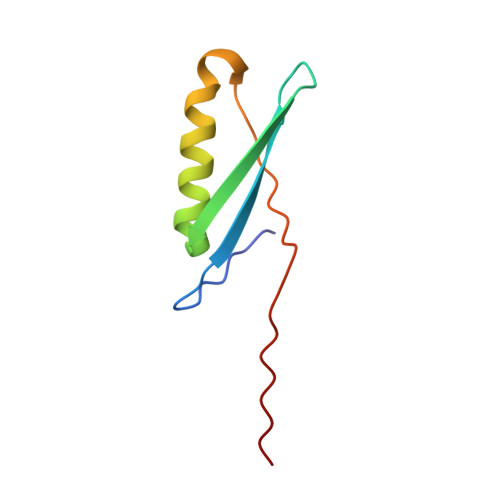
1JML

Conversion of Monomeric Protein L to an Obligate Dimer by Computational Protein Design


Experimental Data Snapshot

  • Method: X-RAY DIFFRACTION
  • Resolution: 1.90 Å
  • R-Value Free: 0.218 
  • R-Value Work: 0.193 

Starting Model: experimental
View more details

wwPDB Validation   3D Report Full Report


This is version 1.6 of the entry. See complete history


Literature

Conversion of monomeric protein L to an obligate dimer by computational protein design.

Kuhlman, B.O'Neill, J.W.Kim, D.E.Zhang, K.Y.Baker, D.

(2001) Proc Natl Acad Sci U S A 98: 10687-10691

  • DOI: https://doi.org/10.1073/pnas.181354398
  • Primary Citation of Related Structures:  
    1JML

  • PubMed Abstract: 

    Protein L consists of a single alpha-helix packed on a four-stranded beta-sheet formed by two symmetrically opposed beta-hairpins. We use a computer-based protein design procedure to stabilize a domain-swapped dimer of protein L in which the second beta-turn straightens and the C-terminal strand inserts into the beta-sheet of the partner. The designed obligate dimer contains three mutations (A52V, N53P, and G55A) and has a dissociation constant of approximately 700 pM, which is comparable to the dissociation constant of many naturally occurring protein dimers. The structure of the dimer has been determined by x-ray crystallography and is close to the in silico model.


  • Organizational Affiliation

    Department of Biochemistry and Howard Hughes Medical Institute, University of Washington, Seattle, WA 98195, USA.


Macromolecules
Find similar proteins by:  (by identity cutoff)  |  3D Structure
Entity ID: 1
MoleculeChains Sequence LengthOrganismDetailsImage
Protein L72Finegoldia magna ATCC 29328Mutation(s): 4 
UniProt
Find proteins for Q51912 (Finegoldia magna)
Explore Q51912 
Go to UniProtKB:  Q51912
Entity Groups  
Sequence Clusters30% Identity50% Identity70% Identity90% Identity95% Identity100% Identity
UniProt GroupQ51912
Sequence Annotations
Expand
  • Reference Sequence
Experimental Data & Validation

Experimental Data

  • Method: X-RAY DIFFRACTION
  • Resolution: 1.90 Å
  • R-Value Free: 0.218 
  • R-Value Work: 0.193 
  • Space Group: C 2 2 21
Unit Cell:
Length ( Å )Angle ( ˚ )
a = 46.762α = 90
b = 76.744β = 90
c = 59.797γ = 90
Software Package:
Software NamePurpose
EPMRphasing
CNSrefinement
SCALEPACKdata scaling

Structure Validation

View Full Validation Report


Entry History 

Deposition Data

Revision History  (Full details and data files)

  • Version 1.0: 2001-10-10
    Type: Initial release
  • Version 1.1: 2008-04-27
    Changes: Version format compliance
  • Version 1.2: 2011-07-13
    Changes: Derived calculations, Source and taxonomy, Version format compliance
  • Version 1.3: 2017-02-01
    Changes: Structure summary
  • Version 1.4: 2017-10-04
    Changes: Refinement description
  • Version 1.5: 2021-10-27
    Changes: Database references, Derived calculations
  • Version 1.6: 2023-08-16
    Changes: Data collection, Refinement description