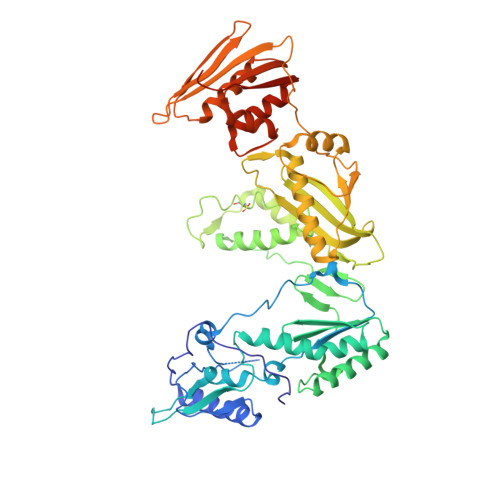
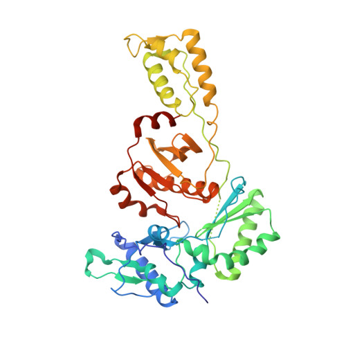
1JLQ

CRYSTAL STRUCTURE OF HIV-1 REVERSE TRANSCRIPTASE IN COMPLEX WITH 739W94


Experimental Data Snapshot

  • Method: X-RAY DIFFRACTION
  • Resolution: 3.00 Å
  • R-Value Free: 0.267 
  • R-Value Work: 0.219 
  • R-Value Observed: 0.219 

wwPDB Validation   3D Report Full Report


Ligand Structure Quality Assessment 


This is version 1.6 of the entry. See complete history


Literature

2-Amino-6-arylsulfonylbenzonitriles as non-nucleoside reverse transcriptase inhibitors of HIV-1.

Chan, J.H.Hong, J.S.Hunter III, R.N.Orr, G.F.Cowan, J.R.Sherman, D.B.Sparks, S.M.Reitter, B.E.Andrews III., C.W.Hazen, R.J.St Clair, M.Boone, L.R.Ferris, R.G.Creech, K.L.Roberts, G.B.Short, S.A.Weaver, K.Ott, R.J.Ren, J.Hopkins, A.Stuart, D.I.Stammers, D.K.

(2001) J Med Chem 44: 1866-1882

  • DOI: https://doi.org/10.1021/jm0004906
  • Primary Citation of Related Structures:  
    1JLQ

  • PubMed Abstract: 

    A series of 2-amino-5-arylthiobenzonitriles (1) was found to be active against HIV-1. Structural modifications led to the sulfoxides (2) and sulfones (3). The sulfoxides generally showed antiviral activity against HIV-1 similar to that of 1. The sulfones, however, were the most potent series of analogues, a number having activity against HIV-1 in the nanomolar range. Structural-activity relationship (SAR) studies suggested that a meta substituent, particularly a meta methyl substituent, invariably increased antiviral activities. However, optimal antiviral activities were manifested by compounds where both meta groups in the arylsulfonyl moiety were substituted and one of the substituents was a methyl group. Such a disubstitution led to compounds 3v, 3w, 3x, and 3y having IC50 values against HIV-1 in the low nanomolar range. When gauged for their broad-spectrum antiviral activity against key non-nucleoside reverse transcriptase inhibitor (NNRTI) related mutants, all the di-meta-substituted sulfones 3u-z and the 2-naphthyl analogue 3ee generally showed single-digit nanomolar activity against the V106A and P236L strains and submicromolar to low nanomolar activity against strains E138K, V108I, and Y188C. However, they showed a lack of activity against the K103N and Y181C mutant viruses. The elucidation of the X-ray crystal structure of the complex of 3v (739W94) in HIV-1 reverse transcriptase showed an overlap in the binding domain when compared with the complex of nevirapine in HIV-1 reverse transcriptase. The X-ray structure allowed for the rationalization of SAR data and potencies of the compounds against the mutants.


  • Organizational Affiliation

    Glaxo Wellcome, Inc., 5 Moore Drive, Research Triangle Park, North Carolina 27709, USA. jc25572@glaxowellcome.com


Macromolecules
Find similar proteins by:  (by identity cutoff)  |  3D Structure
Entity ID: 1
MoleculeChains Sequence LengthOrganismDetailsImage
HIV-1 RT, A-CHAIN560HIV-1 M:B_HXB2RMutation(s): 1 
EC: 2.7.7.49
UniProt
Find proteins for P04585 (Human immunodeficiency virus type 1 group M subtype B (isolate HXB2))
Explore P04585 
Go to UniProtKB:  P04585
Entity Groups  
Sequence Clusters30% Identity50% Identity70% Identity90% Identity95% Identity100% Identity
UniProt GroupP04585
Sequence Annotations
Expand
  • Reference Sequence
Find similar proteins by:  (by identity cutoff)  |  3D Structure
Entity ID: 2
MoleculeChains Sequence LengthOrganismDetailsImage
HIV-1 RT, B-CHAIN440HIV-1 M:B_HXB2RMutation(s): 0 
EC: 2.7.7.49
UniProt
Find proteins for P04585 (Human immunodeficiency virus type 1 group M subtype B (isolate HXB2))
Explore P04585 
Go to UniProtKB:  P04585
Entity Groups  
Sequence Clusters30% Identity50% Identity70% Identity90% Identity95% Identity100% Identity
UniProt GroupP04585
Sequence Annotations
Expand
  • Reference Sequence
Small Molecules
Ligands 1 Unique
IDChains Name / Formula / InChI Key2D Diagram3D Interactions
SBN
Query on SBN

Download Ideal Coordinates CCD File 
C [auth A]2-AMINO-6-(3,5-DIMETHYLPHENYL)SULFONYLBENZONITRILE
C15 H14 N2 O2 S
SWGDXLAZBZDUBR-UHFFFAOYSA-N
Modified Residues  1 Unique
IDChains TypeFormula2D DiagramParent
CSD
Query on CSD
A
L-PEPTIDE LINKINGC3 H7 N O4 SCYS
Binding Affinity Annotations 
IDSourceBinding Affinity
SBN BindingDB:  1JLQ IC50: min: 7, max: 6998 (nM) from 4 assay(s)
PDBBind:  1JLQ IC50: 7 (nM) from 1 assay(s)
Experimental Data & Validation

Experimental Data

  • Method: X-RAY DIFFRACTION
  • Resolution: 3.00 Å
  • R-Value Free: 0.267 
  • R-Value Work: 0.219 
  • R-Value Observed: 0.219 
  • Space Group: P 21 21 21
Unit Cell:
Length ( Å )Angle ( ˚ )
a = 141.1α = 90
b = 110.9β = 90
c = 73.1γ = 90
Software Package:
Software NamePurpose
DENZOdata reduction
SCALEPACKdata scaling
CNSrefinement
CNSphasing

Structure Validation

View Full Validation Report


Ligand Structure Quality Assessment 


Entry History 

Deposition Data

Revision History  (Full details and data files)

  • Version 1.0: 2001-08-22
    Type: Initial release
  • Version 1.1: 2008-04-27
    Changes: Version format compliance
  • Version 1.2: 2011-07-13
    Changes: Version format compliance
  • Version 1.3: 2018-01-24
    Changes: Database references
  • Version 1.4: 2018-01-31
    Changes: Database references
  • Version 1.5: 2022-12-21
    Changes: Database references, Derived calculations
  • Version 1.6: 2024-11-06
    Changes: Data collection, Structure summary