1JL5 | pdb_00001jl5

Novel Molecular Architecture of YopM-a Leucine-rich Effector Protein from Yersinia pestis


Experimental Data Snapshot

  • Method: X-RAY DIFFRACTION
  • Resolution: 2.10 Å
  • R-Value Free: 
    0.240 (Depositor), 0.231 (DCC) 
  • R-Value Work: 
    0.220 (Depositor), 0.221 (DCC) 
  • R-Value Observed: 
    0.220 (Depositor) 

Starting Model: experimental
View more details

wwPDB Validation 3D Report Full Report

Validation slider image for 1JL5

This is version 1.3 of the entry. See complete history

Literature

Unusual molecular architecture of the Yersinia pestis cytotoxin YopM: a leucine-rich repeat protein with the shortest repeating unit.

Evdokimov, A.G.Anderson, D.E.Routzahn, K.M.Waugh, D.S.

(2001) J Mol Biology 312: 807-821

  • DOI: https://doi.org/10.1006/jmbi.2001.4973
  • Primary Citation Related Structures: 
    1G9U, 1JL5

  • PubMed Abstract: 

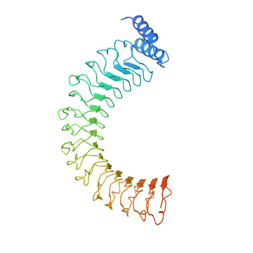
    Many Gram-negative bacterial pathogens employ a contact-dependent (type III) secretion system to deliver effector proteins into the cytosol of animal or plant cells. Collectively, these effectors enable the bacteria to evade the immune response of the infected organism by modulating host-cell functions. YopM, a member of the leucine-rich repeat protein superfamily, is an effector produced by the bubonic plague bacterium, Yersinia pestis, that is essential for virulence. Here, we report crystal structures of YopM at 2.4 and 2.1 A resolution. Among all leucine-rich repeat family members whose atomic coordinates have been reported, the repeating unit of YopM has the least canonical secondary structure. In both crystals, four YopM monomers form a hollow cylinder with an inner diameter of 35 A. The domain that targets YopM for translocation into eukaryotic cells adopts a well-ordered, alpha-helical conformation that packs tightly against the proximal leucine-rich repeat module. A similar alpha-helical domain can be identified in virulence-associated leucine-rich repeat proteins produced by Salmonella typhimurium and Shigella flexneri, and in the conceptual translation products of several open reading frames in Y. pestis.


  • Organizational Affiliation
    • Protein Engineering Section Macromolecular Crystallography Laboratory, National Cancer Institute at Frederick, Frederick, MD 21702-1201, USA. eudokima@ncifcrf.gov

Macromolecule Content 

  • Total Structure Weight: 51.79 kDa 
  • Atom Count: 3,137 
  • Modeled Residue Count: 353 
  • Deposited Residue Count: 454 
  • Unique protein chains: 1

Macromolecules

Find similar proteins by:|  3D Structure
Entity ID: 1
MoleculeChains  Sequence LengthOrganismDetailsImage
outer protein YopM454Yersinia pestisMutation(s): 0 
Gene Names: YopM
UniProt
Find proteins for P17778 (Yersinia pestis)
Explore P17778 
Go to UniProtKB:  P17778
Entity Groups
Sequence Clusters30% Identity50% Identity70% Identity90% Identity95% Identity100% Identity
UniProt GroupP17778
Sequence Annotations
Expand
Reference Sequence

Experimental Data & Validation

Experimental Data

  • Method: X-RAY DIFFRACTION
  • Resolution: 2.10 Å
  • R-Value Free:  0.240 (Depositor), 0.231 (DCC) 
  • R-Value Work:  0.220 (Depositor), 0.221 (DCC) 
  • R-Value Observed: 0.220 (Depositor) 
Space Group: I 41 2 2
Unit Cell:
Length ( Å )Angle ( ˚ )
a = 149.43α = 90
b = 149.43β = 90
c = 191.18γ = 90
Software Package:
Software NamePurpose
AMoREphasing
CNSrefinement
DENZOdata reduction
SCALEPACKdata scaling

Structure Validation

View Full Validation Report


Entry History 

Deposition Data

Revision History  (Full details and data files)

  • Version 1.0: 2001-10-10
    Type: Initial release
  • Version 1.1: 2008-04-27
    Changes: Version format compliance
  • Version 1.2: 2011-07-13
    Changes: Version format compliance
  • Version 1.3: 2023-08-16
    Changes: Data collection, Database references, Derived calculations, Refinement description