The structure of Ap(4)A hydrolase complexed with ATP-MgF(x) reveals the basis of substrate binding.
Fletcher, J.I., Swarbrick, J.D., Maksel, D., Gayler, K.R., Gooley, P.R.(2002) Structure 10: 205-213
- PubMed: 11839306 Search on PubMed
- DOI: https://doi.org/10.1016/s0969-2126(02)00696-2
- Primary Citation Related Structures:
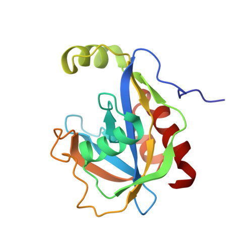
1JKN - PubMed Abstract:
Ap(4)A hydrolases are Nudix enzymes that regulate intracellular dinucleoside polyphosphate concentrations, implicating them in a range of biological events, including heat shock and metabolic stress. We have demonstrated that ATP x MgF(x) can be used to mimic substrates in the binding site of Ap(4)A hydrolase from Lupinus angustifolius and that, unlike previous substrate analogs, it is in slow exchange with the enzyme. The three-dimensional structure of the enzyme complexed with ATP x MgF(x) was solved and shows significant conformational changes. The substrate binding site of L. angustifolius Ap(4)A hydrolase differs markedly from the two previously published Nudix enzymes, ADP-ribose pyrophosphatase and MutT, despite their common fold and the conservation of active site residues. The majority of residues involved in substrate binding are conserved in asymmetrical Ap(4)A hydrolases from pathogenic bacteria, but are absent in their human counterparts, suggesting that it might be possible to generate compounds that target bacterial, but not human, Ap(4)A hydrolases.
- Department of Biochemistry and Molecular Biology, The University of Melbourne, Victoria 3010, Australia.
Organizational Affiliation: