1JID | pdb_00001jid

Human SRP19 in complex with helix 6 of Human SRP RNA


Experimental Data Snapshot

  • Method: X-RAY DIFFRACTION
  • Resolution: 1.80 Å
  • R-Value Free: 
    0.222 (Depositor), 0.212 (DCC) 
  • R-Value Work: 
    0.186 (Depositor), 0.178 (DCC) 

Starting Model: experimental
View more details

wwPDB Validation 3D Report Full Report

Validation slider image for 1JID

This is version 1.4 of the entry. See complete history

Literature

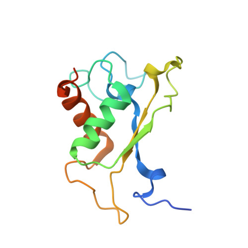
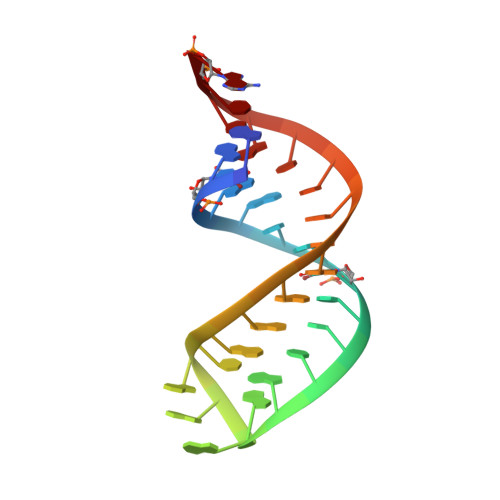
Crystal structure of an early protein-RNA assembly complex of the signal recognition particle.

Wild, K.Sinning, I.Cusack, S.

(2001) Science 294: 598-601

  • DOI: https://doi.org/10.1126/science.1063839
  • Primary Citation Related Structures: 
    1JID

  • PubMed Abstract: 

    The signal recognition particle (SRP) is a universally conserved ribonucleoprotein complex that mediates the cotranslational targeting of secretory and membrane proteins to cellular membranes. A crucial early step in SRP assembly in archaea and eukarya is the binding of protein SRP19 to specific sites on SRP RNA. Here we report the 1.8 angstrom resolution crystal structure of human SRP19 in complex with its primary binding site on helix 6 of SRP RNA, which consists of a stem-loop structure closed by an unusual GGAG tetraloop. Protein-RNA interactions are mediated by the specific recognition of a widened major groove and the tetraloop without any direct protein-base contacts and include a complex network of highly ordered water molecules. A model of the assembly of the SRP core comprising SRP19, SRP54, and SRP RNA based on crystallographic and biochemical data is proposed.


  • Organizational Affiliation
    • Biochemie-Zentrum (BZH), University of Heidelberg, Im Neuenheimer Feld 328, D-69120 Heidelberg, Germany. klemens.wild@bzh.uni-heidelberg.de

Macromolecule Content 

  • Total Structure Weight: 24.47 kDa 
  • Atom Count: 1,828 
  • Modeled Residue Count: 143 
  • Deposited Residue Count: 157 
  • Unique protein chains: 1
  • Unique nucleic acid chains: 1

Macromolecules


Find similar proteins by:|  3D Structure
Entity ID: 2
MoleculeChains  Sequence LengthOrganismDetailsImage
SIGNAL RECOGNITION PARTICLE 19 KDA PROTEINB [auth A]128Homo sapiensMutation(s): 0 
UniProt & NIH Common Fund Data Resources
Find proteins for P09132 (Homo sapiens)
Explore P09132 
Go to UniProtKB:  P09132
PHAROS:  P09132
GTEx:  ENSG00000153037 
Entity Groups
Sequence Clusters30% Identity50% Identity70% Identity90% Identity95% Identity100% Identity
UniProt GroupP09132
Sequence Annotations
Expand
Reference Sequence
Find similar nucleic acids by:  (by identity cutoff) 
Entity ID: 1
MoleculeChains LengthOrganismImage
HELIX 6 OF HUMAN SRP RNAA [auth B]29N/A
Sequence Annotations
Expand
Reference Sequence

Experimental Data & Validation

Experimental Data

  • Method: X-RAY DIFFRACTION
  • Resolution: 1.80 Å
  • R-Value Free:  0.222 (Depositor), 0.212 (DCC) 
  • R-Value Work:  0.186 (Depositor), 0.178 (DCC) 
Space Group: C 2 2 21
Unit Cell:
Length ( Å )Angle ( ˚ )
a = 55.813α = 90
b = 109.782β = 90
c = 89.461γ = 90
Software Package:
Software NamePurpose
CNSrefinement
DENZOdata reduction
SCALEPACKdata scaling
CNSphasing
SHARPphasing

Structure Validation

View Full Validation Report


Entry History 

Deposition Data

Revision History  (Full details and data files)

  • Version 1.0: 2001-10-19
    Type: Initial release
  • Version 1.1: 2008-04-27
    Changes: Version format compliance
  • Version 1.2: 2011-07-13
    Changes: Version format compliance
  • Version 1.3: 2021-10-27
    Changes: Database references, Derived calculations
  • Version 1.4: 2023-08-16
    Changes: Data collection, Refinement description